Case Author(s): J. Philip Moyers, M.D. and Farrokh Dehdashti, M.D. , 10/1/95 . Rating: #D3, #Q4
Diagnosis: Metastatic bronchoalveolar carcinoma
Brief history:
Status post right upper
lobectomy for bronchoalveolar carcinoma.
Images:
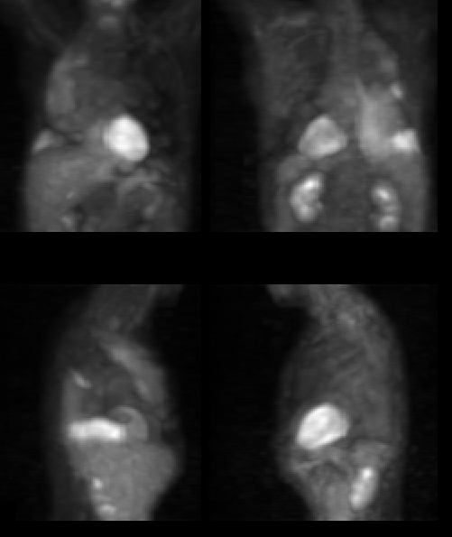
Images from a whole body PET scan
View main image(pt) in a separate image viewer
View second image(xr).
PA chest radiograph
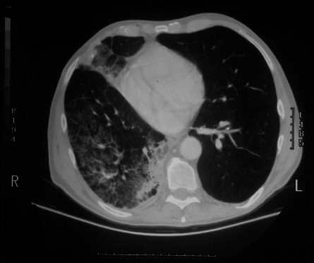
View third image(ct).
Single image from chest CT
Full history/Diagnosis is available below
Diagnosis: Metastatic bronchoalveolar carcinoma
Full history:
66-year old man status post right
upper lobectomy for bronchalveolar carcinoma. The
patient presents with worsening cough and persistent
right lung infiltrate. The patient was being evaluated
for complete right pneumonectomy. This examination
is obtained to evaluate for mediastinal disease, as well
as left lung disease.
Radiopharmaceutical:
14.8 mCi F-18
fluorodeoxyglucose i.v.
Findings:
PET images demonstrated increased FDG accumulation
diffusely throughout the right lung consistent with
the radiographic and CT abnormalities demonstrated
diffusely throughout the right lung. These findings
are consistent with metastatic bronchoalveolar
carcinoma to the right middle and lower lobes.
However, no activity is demonstrated in the left lobe
and mediastinum.
Discussion:
Bronchoalveolar carcinoma
accounts for between 1-20% of pulmonary neoplasms.
The population most affected is middle-aged, with no
predilection for either sex. Interestingly, there is an
increased incidence in patients with scleroderma or
other diseases causing localized parenchymal scarring
or diffuse interstitial fibrosis. Diffuse bilateral
involvement in bronchoalveolar cell carcinoma occurs
late in the disease and is usually spread by the
bronchial tree. Manifestations include both local and
diffuse forms. The local form may grow very slowly
changing little for several years. The diffuse form
simulates an airspace filling disease with air
bronchograms and air broncholograms. A pleural
effusion develops in 8-10% of cases. Uncommon
manifestations are mediastinal adenopathy,
spontaneous pneumothorax, or atelectasis. cavitation
also is uncommon. This disease has part of the
differential of chronic air space disease for which the
following pneumonic may be helpful. TBALLS, T-
tuberculosis. B-bronchalveolar. A-alveolar
proteinosis. L-lipoid pneumonia. L-lymphoma. S-
sarcoidosis.
Although computed tomography and magnetic
resonance imaging have played an important role in
the diagnosis, staging and treatment response of lung
tumors, recent studies have shown limitations with
both techniques. These modalities provide excellent
anatomic information, but not metabolic or
pathophysiologic information of the lesion. For
example, the prospective data from the Multi-
institutional Radiologic Diagnostic Oncology Group
Trial, sponsored by the National Cancer Institute,
showed that in staging non-small cell lung cancer, CT
was only 52% sensitive and 69% specific, whereas MR
imaging was 48% sensitive and 64% specific. PET
imaging with F-18-fluorodeoxyglucose (FDG) has been
shown to be useful for assessing solitary nodules,
mediastinal staging, and assessment of reponse to
therapy on the basis of the differential uptake in non-
neoplastic and malignant lesions. The measurement
of FDG uptake provides an index of glucose
metabolism in tumors, which, in turn, is used to assist
in diagnostic work-up and evaluation of treatment
options.
References:
1) Pare, Fraser. Synopsysis of disease of the chest.
WB Saunders.
2) Abe Y, Matsuzawa T, Fujiwara T, Itoh M,
Fukuda H, Yamaguchi K, Kubota K, Hatazawa J,
Tada M, Ido T, Watanuki S. Clinical assessment
of the therapeutic effects on cancer using 18F-2-
fluoro-2-deoxy-D-glucose and positron emission
tomography: preliminary study of lung cancer.
Int J Radiation Oncology Biol Phys 1990;
19:1005-1010.
3) McLoud TC, Bourgouin PM, Greenberg RW,
Kosiuk JP, Templeton PA, Shepard JO, Moore
EH, Wain JC, Mathisen DJ, Grillo HC.
Bronchogenic carcinoma: analysis of staging in
the mediastinum with CT by correlative lymph
node mapping and sampling. Radiology 1992;
182:319-323.
4) Wahl RL, Quint LE, Greenough RL, Meyer CR,
White RI, Orringer MB. Staging of mediastinal
non-small cell lung cancer with FDG PET, CT,
and fusion images: preliminary prospective
evaluation. Radiology 1994; 191:371-377.
5) Minn H, Zasadny KR, Quint LE, Wahl RL. Lung
cancer: reproducibility of quantitative
measurements for evaluating 2
Followup:
The patient had a sputum cytology
positive for bronchoalveolar carcinoma and at this
time was awaiting right pneumonectomy for
symptomatic relief.
Major teaching point(s):
Abnormal areas of FDG
uptake suggest areas of increased metabolism. These
can be seen in carcinoma such as bronchoalveolar
carcinoma. In this case, diffuse activity throughout
the right lung suggest diffuse involvement by tumor.
The patientąs previous history of right upper
lobectomy for bronchoalveolar carcinoma as well as
the hazy infiltrates demonstrated throughout the
right lung suggests metastatic bronchoalveolar
carcinoma. These findings are confirmed on the PET
study.
ACR Codes and Keywords:
References and General Discussion of PET Tumor Imaging Studies (Anatomic field:Lung, Mediastinum, and Pleura, Category:Neoplasm, Neoplastic-like condition)
Search for similar cases.
Edit this case
Add comments about this case
Read comments about this case
Return to the Teaching File home page.
Case number: pt005
Copyright by Wash U MO