Case Author(s): Charles Pringle, M.D./ F. Dehdashti, M.D. , 10/17/95 . Rating: #D4, #Q4
Diagnosis: Metastatic malignant melanoma
Brief history:
Past history of malignant
melanoma.
Images:

anterior whole body
View main image(pt) in a separate image viewer
View second image(ct).
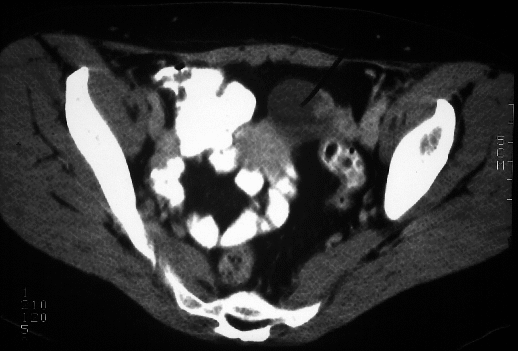
C. T. scan through pelvis
View third image(ct).
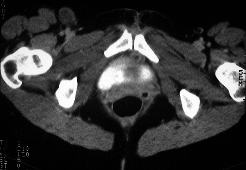
C. T. scan through inguinal region
View fourth image(ct).
C. T. scan (lung windows) through chest
Full history/Diagnosis is available below
Diagnosis: Metastatic malignant melanoma
Full history:
38-year old woman with history
of malignant melanoma eleven years ago. Now
presents with metastases in a right inguinal node.
Evaluate for other areas of metastatic disease.
Findings:
Multiple focal areas of increased
FDG accumulation are present in the thorax,
abdomen, and pelvis. On the anterior whole body
image, the abnormally increased activity within the
left pelvis, right inguinal region, and mediastinum is
seen. These areas are suspicious for metastatic
disease.
Discussion:
None
Followup:
A left salpingo-oophorectomy was
performed, which revealed a hemorrhagic corpus
luteum cyst, paraovarian adhesions, and no evidence
of cancer. Right inguinal node biopsy was positive for
malignant melanoma.
Major teaching point(s):
The findings on the PET
study are significant in that in addition to the
malignancy, the hemorrhagic cystic lesion in the ovary
also showed increased metabolism. The exact
mechanism of increased FDG accumulation in this
benign lesion is unknown.
ACR Codes and Keywords:
References and General Discussion of PET Tumor Imaging Studies (Anatomic field:Vascular and Lymphatic Systems, Category:Misc)
Search for similar cases.
Edit this case
Add comments about this case
Return to the Teaching File home page.
Case number: pt004
Copyright by Wash U MO