Case Author(s): Charles Pringle / Barry Siegel , 7/7/95 . Rating: #D3, #Q4
Diagnosis: Metastatic esophogeal adenocarcinoma
Brief history:
Dysphagia and refux.
Images:
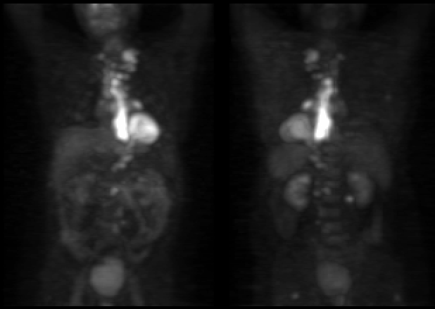
Anterior and posterior reprojection images.
View main image(pt) in a separate image viewer
View second image(fl).
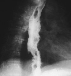
esophogram
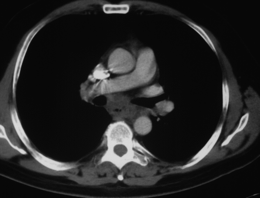
View third image(ct).
Full history/Diagnosis is available below
Diagnosis: Metastatic esophogeal adenocarcinoma
Full history:
44-year old man with newly
diagnosed esophageal carcinoma. This examination
was requested for staging.
Findings:
Increased FDG accumulation
within the mid to distal esophagus is present and
correlates with the patient's primary esophageal
carcinoma. There are multiple focal areas of
increased FDG accumulation within left
supraclavicular, anterior mediastinal, right
paratracheal, left hilar, gastrohepatic-ligament, and
retroperitoneal lymph nodes. There also are several
areas of increased FDG accumulation within the
soft tissues near the tip of the right scapula, the right
paravertebral region at the L2 level, and bilaterally
in the gluteal region at the level of the hips.
Discussion:
Upper GI series obtained on 6-16-95 revealed
a fixed lobulated esophageal mass
extending from approximately the level of the
carina to the gastroesophageal junction. CT scan
obtained 6-29-95 again demonstrated this
esophageal mass and additionally lymphadenopathy
involving the pretracheal, right paratracheal,
aortopulmonary window, subcarinal, and pericardial
regions and, also most likely, the infrahilar region.
These areas correspond to the patient's PET
abnormalities. No definite abnormal CT correlates
were identified for the gastrohepatic or
retroperitoneal lymph nodes or for the soft tissue
lesions identified on the PET study in the region of
the right scapular tip, right paravertebral region, and
bilateral gluteal regions.
ACR Codes and Keywords:
References and General Discussion of PET Tumor Imaging Studies (Anatomic field:Gasterointestinal System, Category:Neoplasm, Neoplastic-like condition)
Search for similar cases.
Edit this case
Add comments about this case
Read comments about this case
Return to the Teaching File home page.
Case number: pt001
Copyright by Wash U MO