
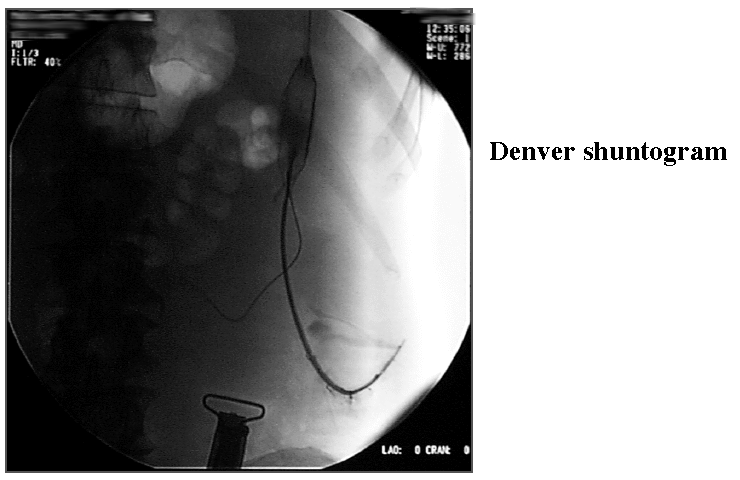
Denver shuntogram
View main image(ps) in a separate image viewer
View second image(fl). Denver shunt contrast injection
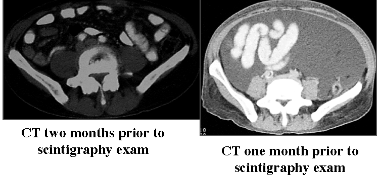
View third image(ct). Computed tomography
Full history/Diagnosis is available below
- Free flow of tracer throughout the peritoneal cavity
- No activity is seen in the blood or lungs. Transmission images (with sheet source behind patient) faintly show expected position of lungs on this image.
2. Computed tomography of chest and abdomen :
- Interval development of massive ascites between the two studies. Chest CT (not shown) also demonstrates numerous pulmonary nodules consistent with metastatic disease.
Intractable ascites is initially treated medically with bedrest, sodium restriction and combinations of diuretics (spironolactone, chlorothiazides, loop diuretics). If ascites is sufficiently severe to cause restricted mobility or respiration and if this is unsuccessful after several weeks, a shunt may be considered.
A variety of other methods have been attempted with limited success. These include repeated paracentesis (this may be complicated by hyponatremia, hypokalemia and hypoproteinemia), ascitic drainage through the bladder, peritoneal glass button (through which ascites may be drained), subcutaneous fistula, hepatoplexy, ileoentrectomoy, thoracic duct drainage and portocaval shunting (Hyde, et al).
Imaging was performed using intraperitoneal Tc-99m MAA, in which a functioning shunt should result in flow to the venous system, with trapping of particles in the lungs. This is preferred over Tc-99m Sulfur-colloid in this setting; the colloid would be taken up in the liver, which might be more easily obscured by the tracer residing in the peritoneum.
References:
Hyde G, Dillon M and Bivins B. Peritoneal Venous Shunting for Ascites: A 15-year perspective. The American Surgeon. 48. 123-127. 1982.
Lund R and Moritz M. Complications of Denver Peritoneovenous Shunting. Arch Surg. 117. 924-928. 1982.
Smith D, Weaver D and Bouwman D. Peritoneovenous shunt (PVS) for malignant ascites. The American Surgeon. 55. 445-449. 1989.
The patient then underwent surgical repair of the venous end of the catheter, and flow was restored.
References and General Discussion of Peritoneal Shunt Scintigraphy (Anatomic field:Gasterointestinal System, Category:Misc)
Return to the Teaching File home page.