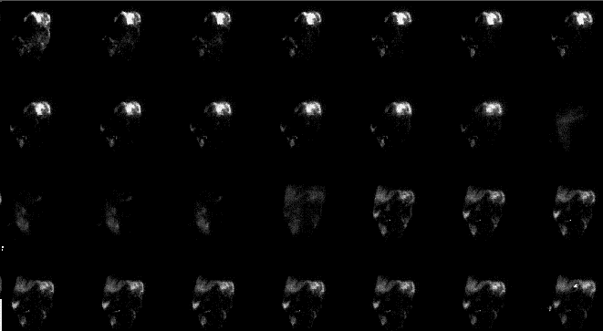
Anterior images centered over the abdomen. What is this study, and why is it being performed?
View main image(ps) in a separate image viewer
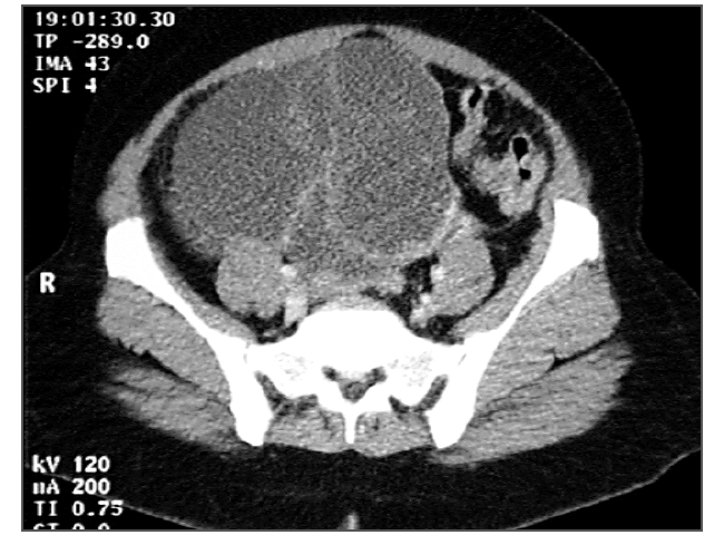
View second image(ct). Computed tomography of abdomen/pelvis
View third image(ps). Selected early and late images from the main image set, shown enlarged and rescaled.
Full history/Diagnosis is available below
- Free flow in the peritoneal cavity through both peritoneal percutaneous peritoneal catheters.
2. Computed tomography of the abdomen and pelvis:
- Large cystic pelvic mass with multiple nodular septations, 14x16x18 cm.
Contrast may also be used to assess the catheter, but is not as easy to interpret and thus entails more risk. Imaging to rule out loculation is best performed with Tc-99m sulfur colloid to assess intraperitoneal distribution. This can then be followed by infusion of chromic phosphate. Approximately 1 liter of ascites fluid was left in place (in one study) when the chromic phosphate is instilled to allow homogeneous distribution.
Note that P-32 is available in two forms: the chromic phosphate intended for intraperitoneal administration, and a soluble form used in much smaller doses for intravenous administration to treat polycythemia vera. Mistaken use of the soluble form for large dose intraperitoneal treatment would likely result in death of the patient, due to diffusion of the tracer into the bloodstream and subsequent bone marrow suppression.
References:
McGowan L. Adjuvant intraperitoneal chromic phosphate therapy in a woman with earlly ovarina carcinoma an pelvic infection with resulting catastrophic complications. Clin Nucl Med. 19, 696-698. 1994.
Tulchinsky M and Eggli D. Intraperitoneal distribution imaging prior to chromic phosphate (P-32) therapy in ovarian cancer patients. Clin Nucl Med. 19, 43-48. 1994
References and General Discussion of Peritoneal Shunt Scintigraphy (Anatomic field:Gasterointestinal System, Category:Neoplasm, Neoplastic-like condition)
Return to the Teaching File home page.