After viewing the image(s), the Full history/Diagnosis is available by using the link here or at the bottom of this page

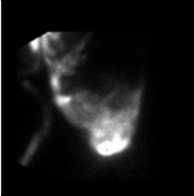
Initial images over the abdomen were obtained.
View main image(ps) in a separate viewing box
View second image(xr). An anterior portable chest radiograph is shown.
View third image(ps). An image over the abdomen and chest with a marker at the sternal notch is shown.
View fourth image(mm). A scintigram of the bandage covering the chest tube insertion is shown.
Full history/Diagnosis is also available
Return to the Teaching File home page.