Case Author(s): Gabriel De Simon, M.D. and Henry Royal, M.D. , . Rating: #D3, #Q4
Diagnosis: Hepatic metastases with central necrosis.
Brief history:
Rectal cancer with right upper quadrant pain.
Images:
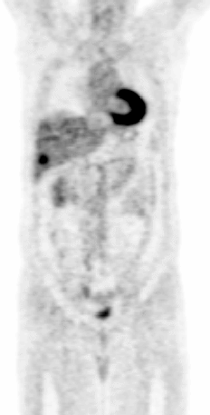
Anterior coronal whole body PET image.
View main image(pt) in a separate image viewer
View second image(pt).
Anterior coronal whole body PET image.
View third image(pe).
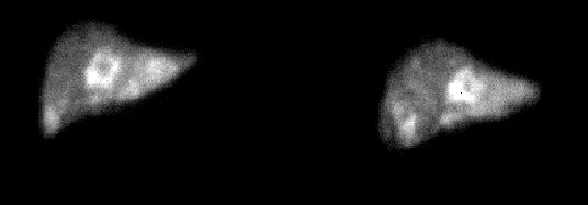
Anterior image of the right upper quadrant.
View fourth image(pe).
Anterior images of the right upper quadrant, 17 days after the initial study.
Full history/Diagnosis is available below
Diagnosis: Hepatic metastases with central necrosis.
Full history:
57 year old male with recently diagnosed rectal cancer. He subsequently had a low anterior resection of the primary rectal cancer and additional resection of a single liver metastasis, followed by chemotherapy.
Radiopharmaceutical:
1. Fluorodeoxyglucose.
2. Technetium 99 macro-aggregated albumin.
Findings:
FDG PET:
Three distinct foci of intensely increased uptake in the liver, two located in the right lobe and the remaining one in the left lobe.
Two small foci of moderately increased activity anteriorly in the left hepatic lobe, questionably additional metastases.
Diffuse mild heterogeneity of hepatic uptake, consistent with previous chemotherapy.
HEPATIC ARTERIAL PERFUSION IMAGING:
Initial study:
Perfusion of the left hepatic lobe predominantly, with only minimal perfusion of the most medial portion of the anterior segment of the right lobe.
Follow up study, 17 days after the initial study:
Marked interval improvement of relative right hepatic lobar perfusion, now approximating that of the left hepatic lobe.
Multiple "ring-like" regions of increased activity surrounding photopenic centers, predominantly in the medial segment of the left hepatic lobe. An additional focus with similar morphological characteristics is present in the inferior right hepatic lobe.
Absence of extra-hepatic activity.
Discussion:
Intra-arterial chemotherapy has been used in the regional treatment of cancer since the early 1960's, delivering high concentration chemotherapy directly to the tumor while minimizing systemic drug exposure and side effects. The rationale for regional chemotherapy is based on the differential blood flow to hepatic tumors. Metastases derive most of their blood supply from the hepatic artery, whereas normal liver cells are supplied predominantly by the portal circulation. Chemotherapy can thus be delivered to the tumor preferentially, minimizing exposure to normal hepatocytes. Hepatic perfusion scintigraphy is indicated in this patient to confirm that the surgically-placed arterial line used for selective infusion of the liver, is optimally positioned. Particulate radiotracers, including Tc 99 MAA used in this study, injected slowly(less than 1 ml/minute), are trapped in hepatic capillaries in proportion to relative blood flow. When chemotherapeutic agents are infused, the goal is to perfuse the entire liver with avoidance of splenic, gastric and bowel perfusion. Shunting of particles through tumor arterio-venous connections results in pulmonary activity, and increases the chances that the patient will develop systemic symptoms from high chemotherapeutic doses due to less hepatic detoxification.
Following confirmation of numerous recurrent liver metastases by both PET imaging and CT, the patient underwent intra-operative ultrasonography, with placement of radiofrequency ablation needles into the centers of several larger metastases. The burns were monitored sonographically. The initial hepatic arterial perfusion study, performed two days after tumor radiofrequency ablation, demonstrates minimal perfusion of the right hepatic lobe compared to the left, attributable to either right hepatic arterial spasm or improper placement of the perfusion catheter. Interval restitution of right hepatic lobar perfusion( follow up study done 17 days later ), without interim catheter adjustment, confirms that vasospasm was indeed the cause of right hepatic lobar hypoperfusion. Numerous, rounded foci are demonstrated, with rims of increased activity likely representing viable tumor, and photopenic centers consistent with necrosis within the metastases. The surrounding increased activity may however, less likely represent hyperemic hepatic tissue following tumor ablation, a distinction which cannot be made solely by hepatic arterial perfusion imaging.
Followup:
The patient presently continues to be on targeted hepatic chemotherapy as an outpatient, and is relatively asymptomatic, with resolution of the upper quadrantal pain. He continues to see the referring physician regularly.
Major teaching point(s):
1. Indications and rationale of targeted selective hepatic chemotherapy.
2. Slow administration of Tc 99 MAA during hepatic arterial scintigraphy allows definition of the pattern of drug perfusion.
3. Extrahepatic perfusion is predictive of severe systemic symptoms following high dose targeted chemotherapy.
ACR Codes and Keywords:
References and General Discussion of Perfusion (only) Scintigraphy (Anatomic field:Gasterointestinal System, Category:Neoplasm, Neoplastic-like condition)
Search for similar cases.
Edit this case
Add comments about this case
Return to the Teaching File home page.
Case number: pe007
Copyright by Wash U MO