Case Author(s): Scott Winner, M.D. and Henry Royal, M.D. , 2/14/97 . Rating: #D3, #Q4
Diagnosis: Malfunctioning photomultiplier tube
Brief history:
The patient presented
with dyspnea and tachypnea
Images:

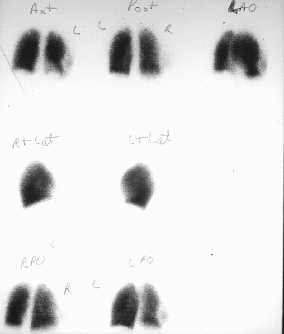
Pulmonary perfusion images
View main image(pe) in a separate image viewer
View second image(pe).
Flood source field uniformity image
Full history/Diagnosis is available below
Diagnosis: Malfunctioning photomultiplier tube
Full history:
As above.
Radiopharmaceutical:
Tc-99m MAA and
Cobalt-57 flood source
Findings:
On the anterior images, there
appears to be a perfusion defect in the lateral
aspect of the left lower lobe. On the left
anterior oblique view, the perfusion defect has
not changed in shape and now appears to be in
the posterior aspect of the left lower lobe. On
the LPO view, the defect appears to be in the
posterior aspect of the right lower lobe. Note
also that the defect always appears in the same
location within the field of view.
Discussion:
The defect seen on the
pulmonary perfusion study was an artifact. A field uniformity image was acquired
and demonstrated a circular region of
decreased activity in the periphery of the field
of view. This appearance is typical for
malfunction of a photomultiplier tube. Each
photomultiplier tube is connected to a
preamplifier and, if the preamplifier
malfunctions, the same round defect will be
seen on the field uniformity image. Because
relatively small changes in field uniformity can
alter the interpretation of clinical studies, it is
imperative to perform field uniformity tests
each day that the camera is used. Other causes
of nonuniformity include fracture of the
crystal, collimator defects, dirt on a photographic lens, and
an asymmetric energy window. All of these causes of nonuniformity are readily distinguishable from an artifact from a malfunctioning PM tube.
Followup:
None
Major teaching point(s):
None
Differential Diagnosis List
See Discussion
ACR Codes and Keywords:
- General ACR code: 61
- Lung, Mediastinum, and Pleura:
6.12171 "Lung perfusion study"
References and General Discussion of Perfusion (only) Scintigraphy (Anatomic field:Lung, Mediastinum, and Pleura, Category:Normal, Technique, Congenital Anomaly)
Search for similar cases.
Edit this case
Add comments about this case
Return to the Teaching File home page.
Case number: pe006
Copyright by Wash U MO

