
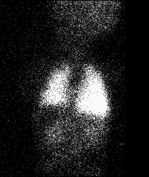
Posterior whole-body image obtained after injection of Tc-99m MAA.
View main image(pe) in a separate image viewer
View second image(pe). Posterior view of the chest and abdomen.
Full history/Diagnosis is available below
To quantify the right-to-left shunt, regions of interest were drawn around the entire body and then, separately, around the lungs to determine the counts in these regions. The counts distributed in the systemic circulatory bed is determined by subtracting the counts in the pulmonary region from the counts in the whole-body region. Based on this study, the right-to-left intracardiac shunt was determined to be 36% (cardiac catheterization with oximetry determined the right-to-left intracardiac shunt to be 33%). "Normal" values determined scintigraphically are 1-10%. This apparent "normal" right-to-left shunt reflects the presence of small particles in the Tc-99m MAA preparation (which are accumulated by phagocytic cells of the reticuloendothelial system, as well as free Tc-99m pertechnetate or other Tc-99m oxides in the radiopharmaceutical preparation or released by breakdown of Tc-99m MAA particles. The finding of activity in the parenchyma of the brain is a definitive sign of a right-to-left shunt rather than excessive free Tc-99m.
Gates et al.(using posterior scintiphotographs) and Lin et al. (using a whole-body profile device) employed this method to quantify right-to-left shunts. One theoretical difficulty is that activity in the lungs and in the remainder of the body may be detected with different sensitivities caused by differences in attenuation. Correction of activity in the pulmonary region of interest for chest wall and mediastinal activity is another methodological difficulty with this technique.
Reference: Pediatric Nuclear Medicine. Treves, S. Second edition. Springer-Verlag, NY, 1995.
References and General Discussion of Perfusion (only) Scintigraphy (Anatomic field:Lung, Mediastinum, and Pleura, Category:Normal, Technique, Congenital Anomaly)
Return to the Teaching File home page.