Case Author(s): Scott Winner, M.D. and Farrokh Dehdashti, M.D. , 9/27/96 . Rating: #D3, #Q3
Diagnosis: Small systemic right to left shunt
Brief history:
46-year old woman with
Weber-Christian disease.
Images:

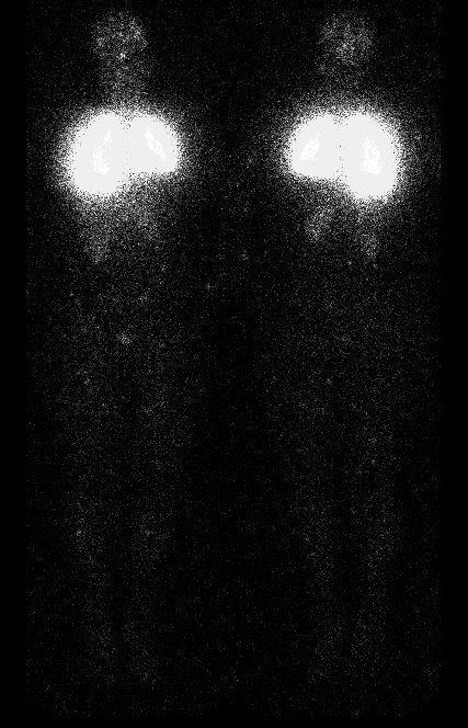
Anterior and posterior whole body images
View main image(pe) in a separate image viewer
View second image(pe).
Anterior and posterior perfusion images
Full history/Diagnosis is available below
Diagnosis: Small systemic right to left shunt
Full history:
46-year old woman with
chronic pulmonary disease, a history of
pulmonary embolism (April, 1996) and Weber-
Christian disease.
Radiopharmaceutical:
11.0 mCi Xe-133
gas by inhalation and 4.3 mCi Tc-99m MAA i.v.
Findings:
The comparison chest radiograph
demonstrates a pacemaker projecting over the right
hemithorax with elevation of the right
hemidiaphragm. Xe-133 ventilation images
demonstrate normal ventilation with the exception
of a defect in the left lung base on the first breath
image. On the washout images, there is retention
of Xe-133 in this area. The perfusion images are
normal; however, a defect related to the pacemaker
is seen projecting on the anterior right lung. In
addition to standard ventilation-perfusion images,
whole-body anterior and posterior views were
obtained, which showed extrapulmonary activity
due to the patient1s known right-to-left shunt.
Quantitative analysis revealed an approximately 9%
systemic right-to-left shunt.
Discussion:
Weber-Christian disease is a chronic disorder characterized by relapsing
febrile episodes and systemic nodular panniculitis (nodular relapsing fat
necrosis). Recurrent crops of tender erythematous nodules may be
accompanied by abdominal pain and fat necrosis in bone marrow, lungs, and other
organs. A variety of distinctive disease entities, such as systemic
lupus erythematosus (SLE), pancreatic disease, alpha-1-antitrypsin
disease, lymphoproliferative neoplasia, infections, or trauma are
associated with chronic panniculitis. The cause of this disease is due
to the effect of pancreatic enzymes released into the circulation on
fatty tissues. The accurate diagnosis of panniculitis requires an
adequate deep skin biopsy showing inflammation of the subcutaneous
layers. The febrile episodes and skin lesions responded dramatically
with the use of oral corticosteroids.
There are two approaches for the quantfication of right-to-left shunt.
One is the angiographic technique using Technetium-99m (Tc-99m) as
pertechnetate or other forms. However, this technique is too cumbersome.
Second is the technique using Tc-99m macroaggregated albumin (MAA).
Whole body images will be performed and the activity in the whole body
will be measured and compared with that in the lungs. In this patient
with a known right to left shunt, scintigraphy with Tc-99m MAA was used
to quantify the shunt. Normally, pulmonary capillaries should trap
nearly all of the Tc-99m MAA on the first pass through the lungs.
However, there is normally a small amount (approximately 4%) of
intravenously injected particles that can cross over to the systemic
circulation (physiologic shunting) but is not visible on routine images.
Note that if the radiopharmaceutical is not freshly prepared, or if
there is a delay after injection prior to imaging, the fraction of
extrapulmonary activitiy in normal patients can be higher due to
breakdown of the Tc-99m MAA.
In this patient with Weber-Christian disease, the calculated value of
9% likely represents a small right to left shunt.
The percent shunt is calculated as follows:
(Total body counts - pulmonary counts)/Total body counts x100
ACR Codes and Keywords:
- General ACR code: 51
- Heart and Great Vessels:
5.12176 "Cardiac shunt quantification"
References and General Discussion of Perfusion (only) Scintigraphy (Anatomic field:Heart and Great Vessels, Category:Normal, Technique, Congenital Anomaly)
Search for similar cases.
Edit this case
Add comments about this case
Read comments about this case
Return to the Teaching File home page.
Case number: pe004
Copyright by Wash U MO

