Case Author(s): M.Roarke, M.D., T. Miller, M.D., Ph.D. , 10/6/95 . Rating: #D2, #Q3
Diagnosis: Iatrogenic occlusion left pulmonary artery, right lower lobe pulmonary artery stenosis.
Brief history:
6 year old status post prior repair of Tetralogy of Fallot.
Images:

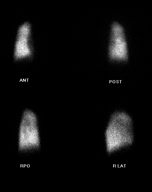
Perfusion lung images, Tc99m MAA
View main image(pe) in a separate image viewer
View second image(an).
Right middle lobe pulmonary artery injection, pulmonary angiogram.
View third image(an).
Right lower lobe pulmonary artery injection, pulmonary angiogram.
Full history/Diagnosis is available below
Diagnosis: Iatrogenic occlusion left pulmonary artery, right lower lobe pulmonary artery stenosis.
Full history:
This is a 6 year-old girl with a history of complete tetralogy of Fallot repair at an outside hospital. The left pulmonary artery was presumably injured during the surgery and, as a consequence, all pulmonary blood flow was directed to the right lung. This examination was requested to quantify the blood flow to the upper lobe, compared to the lower lobe, given the evidence of pulmonary hypertension identified during pulmonary arteriography.
Radiopharmaceutical:
0.96 mCi Tc-99m MAA
Findings:
The perfusion images reveal essentially no perfusion to the left lung, consistent with the known left pulmonary arterial injury. The right upper and middle lobes receive 30% of total flow while the lower lobe receives 70%. The pulmonary arteriogram revealed markedly enlarged and tortuous right upper and middle lobe pulmonary arterial branches consistent with the history of pulmonary hypertension. Additionally, there was a stenosis of the right lower lobe pulmonary artery and normal vascularity of the right lower lobe. The chest radiograph showed enlarged right pulmonary arteries.
Discussion:
The right lower lobe pulmonary arterial stenosis caused preferential blood flow and consequent pulmonary hypertension in the right middle and upper lobes, but protected the right lower lobe pulmoanry vasculature. The pulmonary hypertension led to a disproportionate percentage of pulmonary flow to the right lower lobe.
Differential Diagnosis List
The differential diagnosis for unilateral absent pulmonary perfusion is extensive, but the most common causes include bronchogenic carcinoma, congenital haert disease following Blalock-Taussig shunt or tetralogy of Fallot repair, pneumonectomy, pulmonary embolism, Swyer-James syndrome, and fibrosing mediastinitis (particularly in the Midwest). The age of the patient, clinical presentation, and past medical or surgical history are often helpful in limiting the differential diagnosis.
ACR Codes and Keywords:
References and General Discussion of Perfusion (only) Scintigraphy (Anatomic field:Lung, Mediastinum, and Pleura, Category:Misc)
Search for similar cases.
Edit this case
Add comments about this case
Read comments about this case
Return to the Teaching File home page.
Case number: pe003
Copyright by Wash U MO

