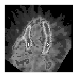

C-11 acetate cardiac PET image
View main image(pc) in a separate image viewer
View second image(pc). C-11 acetate cardiac PET quantitative data
View third image(mi). Myocardial perfusion study
Full history/Diagnosis is available below
- Markedly reduced perfusion and borderline oxidative metabolism involving the apex.
- Normal perfusion and oxidative metabolism elsewhere.
Myocardial perfusion imaging:
- Moderate anteroseptal myocardial infarction with a small area of ischemia
- Moderate-sized inferoseptal myocardial infarction with a small area of ischemia
Several approaches have been used. Flow may be assessed with N-13 ammonia. Areas of low perfusion can then be further assessed with the glucose analog, F-18 fluorodeoxyglucose (FDG). Low flow and low glucose metabolism is consistent with scar (perfusion-metabolism match). Low flow with normal or only slightly low glucose metabolism (perfusion-metabolism mismatch) is consistent with ischemia with viable myocardium. In a tyical scenario, an area of myocardium shows lack of uptake on rest and stress on standard single photon myocardial imaging. This may represent myocardium that is infarcted, hibernating or stunned. PET examination can help make the distinction. If glucose metabolism is intact, or nearly intact, the myocardium is not infarcted. A problem with using FDG to assess viability is that the heart may use glucose or short-chain fatty acids for fuel and therefore glucose utilization is variable. The patient is given a glucose load to encourage use of glucose by the heart. This is in distinction to oncology FDG PET imaging in which the patient is fasted for a minimum of four hours (to prevent high muscle and myocardium uptake that results from insulin release). These physiologic maneuvers are only partially successful, however (for unknown reasons).
Alternatively, short-chain fatty acid metabolism may be ascertained with C-11 palmitate. The clearance rate (due to efflux of C-11 carbon dioxide) is correlated with oxidation. However, the correlation is not strong due to back-diffusion of C-11 palmitate that is not metabolized. C-11 acetate may be used to assess flow with immediate post-injection imaging and to assess oxidative metabolism by performing sequential imaging with calculation of the clearance rate from the various myocardial walls. Clearance rate (reflected by the rate constant of the first phase of C-11 clearance, k1) is correlated with oxidative metabolism (the C-11 is carried away in C-11 carbon dioxide). An advantage of this method is that C-11 acetate assessment of oxidative metabolism is independent of fuel type used (short-chain fatty acids or glucose). It is more accurate than N-13 ammonia / F-18 FDG PET.
Other PET technics that have been used include uptake and retention of Rb-82 and O-15 water-perfusion index (PTI).
References:
Beller G.A. Clinical Nuclear Cardiology. W.B. Saunders Company. 1995.
References and General Discussion of PET Cardiac Imaging Studies (Anatomic field:Heart and Great Vessels, Category:Misc)
Return to the Teaching File home page.