Case Author(s): Richard Held MD, and Jerry Wallis MD , 6/16/06 . Rating: #D4, #Q5
Diagnosis: Alzheimer's Disease
Brief history:
Woman in her mid 30s for evaluation of decreased mental status and level of functioning over two years.
Images:
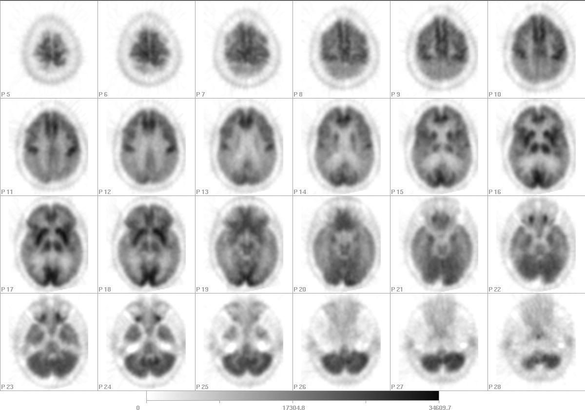
Axial F-18 FDG Brain PET
View main image(pb) in a separate image viewer
View second image(pb).
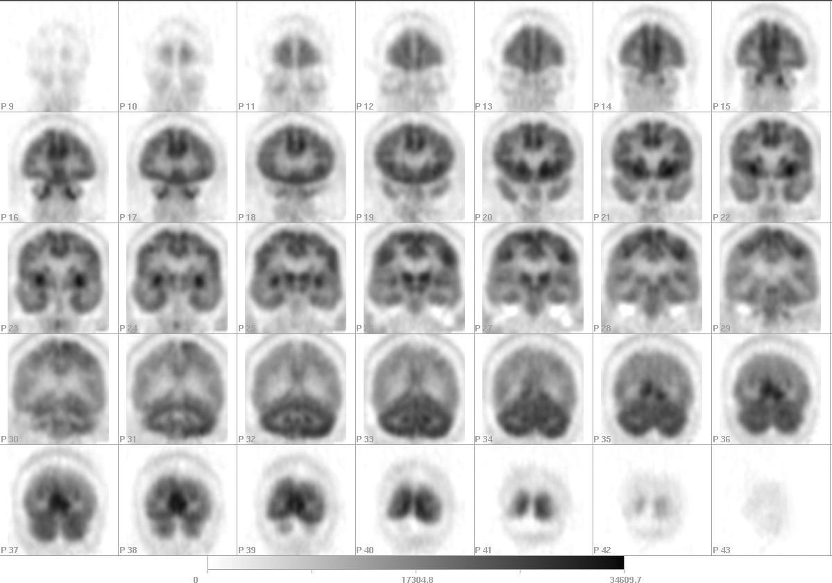
Coronal F-18 FDG Brain PET
Full history/Diagnosis is available below
Diagnosis: Alzheimer's Disease
Full history:
This is a female in her mid 30s who was referred for evaluation of decreased mental status and level of functioning over the past approximately two years.
Initially, the patient was noted to have increasing difficulties performing routine tasks, having difficulty being able to drive to specified locations and getting lost, and who lost her job secondary to inability to be able to perform the specified tasks.
Further workup with EEG at the time demonstrated no EEG abnormalities. The patient's mental status continued to deteriorate over the next 8 months with the patient unable to dress herself properly, often having her clothes on backwards or inside out. Within another 3 months, the patient was unable to read or follow directions, with frequent confusion. At best was able to follow one and maybe two step very simple commands.
The patient underwent repeat MRI scanning 6 months later, demonstrating cortical atrophy. The patient was then referred to Neurology for further evaluation. Basic laboratory studies obtained were normal and Huntington gene testing also was performed which was reported to be normal. The patient presented here for full neurologic evaluation of what appears to be early-onset dementia of a rapidly progressive type,and a PET scan was ordered.
Radiopharmaceutical:
F-18 FDG
Findings:
There is mildly diminished metabolism involving the bilateral frontal lobes. There is also a moderately to marked degree of diminished metabolism involving both the temporal and parietal lobes bilaterally. There is preservation of metabolic activity in the basal ganglia, cerebellum, sensory-motor region, and occipital cortex. There is relatively preserved anterior cingulate gyrus metabolism.
Discussion:
Alzheimer's Disease has a very characteristic pattern of symmetric temporo-parietal lobe hypometabolism, with relative sparing of the sensory motor cortex, and occipital lobes and basal ganglia, as in this case.
Alzheimer's Dementia is one of few Medicare approved indications for Brain PET scanning, and then only after careful screening, documentation, and exclusion of other causes of dementia.
References:
J Nucl Med. 2000 Nov;41(11):1920-8
Movement Disorder, Vol. 16, No. 6, 2001, pp. 1014–1022
Differential Diagnosis List
The pattern of decreased cortical uptake most severe in the temporal and parietal regions with sparing of the basal ganglia is most consistent with Alzheimer's disease. Creutzfeldt-Jacob disease and Parkinson's disease may sometimes give a similar pattern, and should also be considered if this is compatible with the clinical exam.
ACR Codes and Keywords:
References and General Discussion of PET Brain (Nontumor) Imaging Studies (Anatomic field:Skull and Contents, Category:Organ specific)
Search for similar cases.
Edit this case
Add comments about this case
Return to the Teaching File home page.
Case number: pb014
Copyright by Wash U MO