Case Author(s): Zhiyun Yang, M.D. and Henry Royal, M.D. , 09/03/04 . Rating: #D2, #Q3
Diagnosis: Metastatic carcinoma
Brief history:
A 40 year-old woman presented with the acute onset of right-sided weakness and mental status change.
Images:
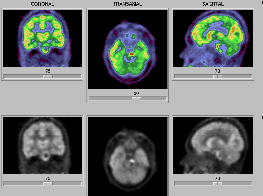
FDG PET brain
View main image(pb) in a separate image viewer
View second image(pb).
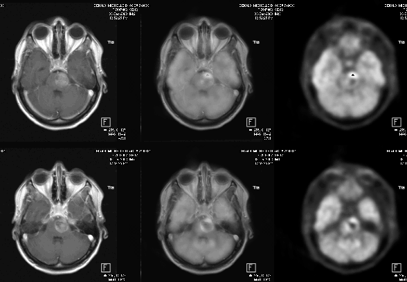
Fusion images of the PET and MRI brain scans
View third image(mr).
MRI brain
View fourth image(pt).
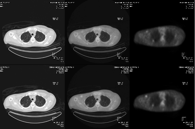
PET-CT images of the chest
Full history/Diagnosis is available below
Diagnosis: Metastatic carcinoma
Full history:
A 40 year old woman developed numbness of her right arm and leg in October, 2003. An MRI of the brain dated 12-3-03 demonstrated a large mass lesion in the pons which has increased in size compared to the prior MRI study dated 11-17-03. MRI brain of 11-17-03 demonstrated a heterogeneous lesion in the pons, which contains blood products. The differential diagnosis includes a cavernoma or a brain stem glioma which has hemorrhaged.
Radiopharmaceutical:
14.7 mCi F-18 Fluorodeoxyglucose i.v.
Findings:
FDG Brain PET images demonstrated a large mass lesion in the pons with centrally decreased activity and a surrounding rim having increased FDG uptake, most consistent with a brain lesion with necrosis or hemorrhage centrally.ĀĀ
Tumor PET images of the whole body demonstrated focally increased FDG uptake in the right upper lobe, which correlates with a right upper lobe cavitary lesion. This cavitary lesion is relatively thin walled, and this likely accounts for the fact that the uptake is relatively mild. This right upper lobe lesion is most consistent with malignancy, although an inflammatory cavitary lesion could have the same appearance.
Discussion:
Clinical indications for FDG PET brain imaging include 1. Differentiating radiation necrosis from a recurrent tumor; 2. characterizing the grade of a primary brain tumor
Followup:
A biopsy of the brain stem lesion revealed poorly differentiated metastatic carcinoma.
Differential Diagnosis List
Brainstem glioma, metastasis, basilar artery aneurysm
ACR Codes and Keywords:
References and General Discussion of PET Brain (Nontumor) Imaging Studies (Anatomic field:Skull and Contents, Category:Neoplasm, Neoplastic-like condition)
Search for similar cases.
Edit this case
Add comments about this case
Return to the Teaching File home page.
Case number: pb013
Copyright by Wash U MO