Case Author(s): Mark Fister, M.D. and Henry D. Royal , 9/20/00 . Rating: #D2, #Q4
Diagnosis: High-grade recurrence of anaplastic astrocytoma.
Brief history:
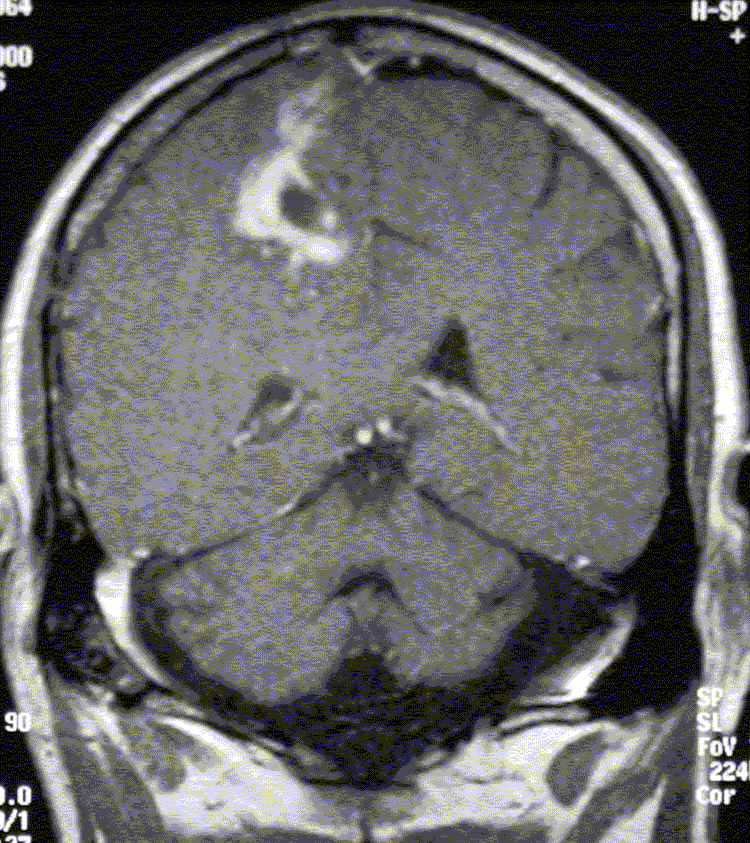
26 year-old man with a history of right parietal anaplastic astrocytoma originally resected 14 years ago, with radiation completed 13 years ago, presents following chemotherapy completed almost 1 year ago with an enlarging mixed solid and cystic enhancing mass in the resection bed by MRI.
Images:

Coronal contrast-enhanced MR image.
View main image(mr) in a separate image viewer
View second image(pb).
Multiplanar F-18 FDG image.
Full history/Diagnosis is available below
Diagnosis: High-grade recurrence of anaplastic astrocytoma.
Full history:
26 year-old man with a history of right parietal anaplastic astrocytoma originally resected 14 years ago, with radiation completed 13 years ago, presents following chemotherapy completed almost 1 year ago with an enlarging mixed solid and cystic enhancing mass in the resection bed by MRI. The patient denies any acute or progressive neurologic symptoms.
Radiopharmaceutical:
F-18 Fluorodeoxyglucose, i.v.
Findings:
F-18 FDG PET reveals a thick, lobulated, irregular rim of intensely increased activity surrounding a central photopenic region in the right frontoparietal region at the site of prior surgery. This correlates well with the mixed cystic and solid enhancing lesion identified on MRI.
Discussion:
The mainstay of therapy for high grade gliomas remains excision followed by radiation therapy. Followup CT or MRI often demonstrate contrast-enhancing areas at the site of prior resection which can be caused by either radiation necrosis or recurrent tumor. Unfortunately, anatomorphometric imaging techniques, such as CT and MRI, cannot reliably differentiate between the two. Metabolic imaging, on the other hand, can. Recurrent high-grade tumor exhibits greater FDG uptake than radionecrosis. A review of the literature by Kuwert and Delbeke reveal an accuracy of FDG-PET for distinguishing between radionecrosis and high-grade recurrence from 71-100%. Moreover, FDG uptake has prognostic significance by virtue of its inverse correlation with survival rate.
Reference: Kuwert T, and Delbeke D. “Brain Tumors” in PET in Clinical Oncology Eds. Coleman RE, and Wieler HJ. Steinkopff Verlag, Darmstadt 2000, pp.125-138.
Followup:
The patient is currently being evaluated for re-excision. If ineligible, a different chemotherapy regimen will be initiated.
Major teaching point(s):
PET is useful for confirming primary brain tumor recurrence and can help differentiate radiation necrosis from viable tumor.
Differential Diagnosis List
1. High-grade tumor recurrence.
2. Brain abscess.
ACR Codes and Keywords:
References and General Discussion of PET Brain (Nontumor) Imaging Studies (Anatomic field:Skull and Contents, Category:Neoplasm, Neoplastic-like condition)
Search for similar cases.
Edit this case
Add comments about this case
Return to the Teaching File home page.
Case number: pb010
Copyright by Wash U MO

