
After viewing the image(s), the Full history/Diagnosis is available by using the link here or at the bottom of this page
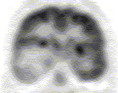
Axial FDG-PET images of the brain are shown
View main image(pb) in a separate viewing box
View second image(pb). A coronal FDG-PET image through the temporal lobes is shown
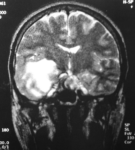
View third image(mr). A sample T2-weighted coronal MRI image through the temporal lobes is shown
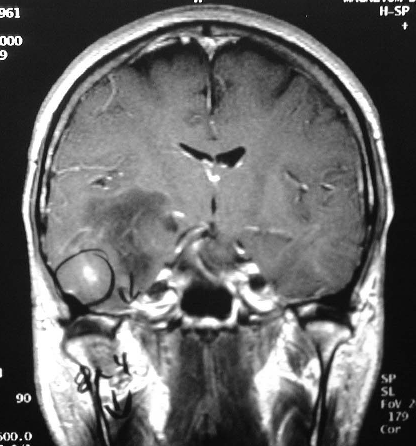
View fourth image(mr). A sample T1-weighted gadolinium-enhanced coronal image through the temporal lobes is shown
Full history/Diagnosis is also available
Return to the Teaching File home page.