Case Author(s): Daniel Appelbaum, MD and Tom R. Miller, MD, PhD , 5/9/00 . Rating: #D3, #Q4
Diagnosis: CNS toxoplasmosis
Brief history:
45 year-old male with mental status changes.
Images:
Selected axial images are shown. What is your differential diagnosis? What can you do next to help differentiate benign from malignant etiologies?
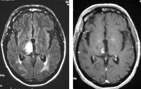
View main image(mr) in a separate image viewer
View second image(pb).
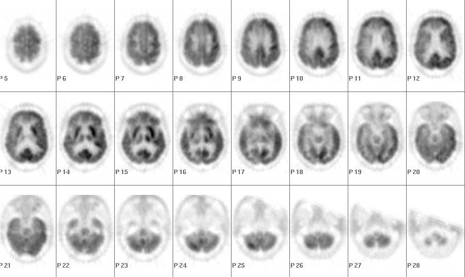
Axial images from the brain PET exam.
View third image(pb).
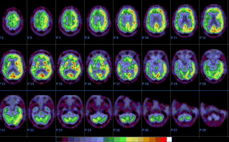
Same axial PET images, for those who prefer color.
View fourth image(pb).
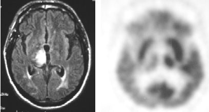
Selected PET and MR images from the same axial level.
Full history/Diagnosis is available below
Diagnosis: CNS toxoplasmosis
Full history:
This 45 year-old HIV-positive male presented with mental status changes. He has a history of prior CNS toxoplasmosis. A brain MR was obtained. A PET examination was then performed for further evaluation.
Radiopharmaceutical:
10.5 mCi F-18 Fluorodeoxyglucose i.v.
Findings:
The MRI from 1/6/00 demonstrates increased signal in the region of the right thalamus. Abnormal enhancement with gadolinium is also present in this location. The brain PET images demonstrate focally decreased FDG activity in the right thalamus. There is also widespread mildly decreased activity of the right cerebral hemisphere, although this spares most of the pre-frontal and primary visual cortices. In addition, mild diffusely decreased activity of the left cerebellum is noted.
Discussion:
For focal enhancing brain lesions in the AIDS population, two primary diagnoses to consider are CNS lymphoma and toxoplasmosis. They can appear indistinguishable on MR, though PET can often differentiate the two. While CNS lymphoma is metabolically very active and thus demonstrates increased FDG uptake (though it may only be equal to or slightly more intense than the prominent activity of normal grey matter), CNS toxoplasmosis is typically more indolent, with only very modest FDG accumulation.
The mild decreased activity involving much of the ipsilateral cerbrum reflects associated mild hypometabolism of this hemisphere. The left cerebellar decreased activity is due to crossed cerebellar diaschisis, a phenomenom often seen with contralateral cerebral pathology.
Followup:
The patient was managed with anti-toxoplasmosis drug therapy with subsequent improvement in symptoms.
ACR Codes and Keywords:
References and General Discussion of PET Brain (Nontumor) Imaging Studies (Anatomic field:Skull and Contents, Category:Inflammation,Infection)
Search for similar cases.
Edit this case
Add comments about this case
Return to the Teaching File home page.
Case number: pb006
Copyright by Wash U MO