Case Author(s): Daniel Appelbaum, MD and Farrokh Dehdashti, MD , 5/8/00 . Rating: #D3, #Q4
Diagnosis: CNS lymphoma
Brief history:
29 year old HIV positive female with a past history of CNS toxoplasmosis now presents with new onset seizures.
Images:

Selected axial and coronal MR images are shown. What is your differential diagnosis? How could you further distinguish between benign and malignant etiologies?
View main image(mr) in a separate image viewer
View second image(pb).
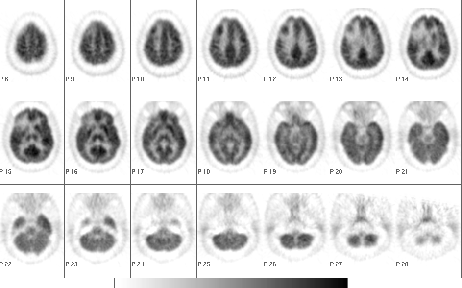
Axial images from a brain PET study.
View third image(pb).
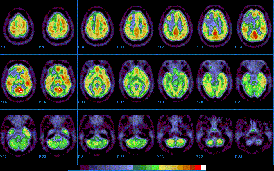
Same axial images, for those who prefer color-scale.
View fourth image(mc).
Corresponding MR and PET images.
Full history/Diagnosis is available below
Diagnosis: CNS lymphoma
Full history:
This is a 29 year old female, HIV positive, who presented with new onset seizures. She had been diagnosed with CNS toxoplasmosis several years ago which involved primarily the deep white matter and basal ganglia. A recent brain MR now demonstrated a new enhancing lesion near the periphery of the right frontal lobe. Given her history, toxoplasmosis recurrence was considered likely. However, given the MR appearance, CNS lymphoma also figured prominently into the differential. PET was obtained to help distinguish between the two possibilities.
Radiopharmaceutical:
10.0 mCi F-18 fluorodeoxyglucose iv.
Findings:
The brain MR demonstrates an enhancing mass in the right frontal lobe with surrounding vasogenic edema. The brain PET demonstrates FDG activity corresponding exactly to the mass lesion. The degree of activity is equal to or slightly greater than the normal grey matter. In addition, a large area of decreased activity surrounds this lesion, which corresponds to edma seen on MRI. Also, there is slightly decreased activity diffusely throughout the left cerebellar hemisphere.
Discussion:
As is seen with lymphoma elsewhere, CNS lymphoma demonstrates increased metabolic activity and thus increased FDG uptake on PET. The lesion is quite metabolically active, the uptake seen in CNS lymphoma may be equal or greater than normal gray matter. The surrounding hypometabolism corresponds to the vasogenic edema. However, FDG uptake in toxoplasmosis is significantly less intense than normal gray matter (or CNS lymphoma). Note, however, that available literature on PET imaging of toxoplasmosis to date involves patients receiving empiric treatment for toxoplasmosis (as they often are). The decreased activity seen in the left cerebellar hemisphere is compatible with slight crossed cerebellar diaschisis, a phenomenon commonly seen with contralateral cerebral cortical pathology.
References:
1. Hoffman et al., J Nucl Med 1993; 34:567-575.
2. Villringer et al., J Comput Assist Tomogr 1995; 19:532-536.
Followup:
The patient had biopsy proven CNS lymphoma.
Major teaching point(s):
FDG-PET is very useful to differentiate CNS lymphoma from toxoplasmosis when MRI is indeterminate.
ACR Codes and Keywords:
References and General Discussion of PET Brain (Nontumor) Imaging Studies (Anatomic field:Skull and Contents, Category:Neoplasm, Neoplastic-like condition)
Search for similar cases.
Edit this case
Add comments about this case
Return to the Teaching File home page.
Case number: pb005
Copyright by Wash U MO