FDG PET scan
View main image(pb) in a separate image viewer
View second image(pb). Repeat FDG PET scan performed 5 weeks later.
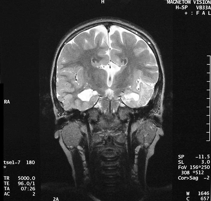
View third image(mr). Brain MR (T2-Weighted Coronal Image)
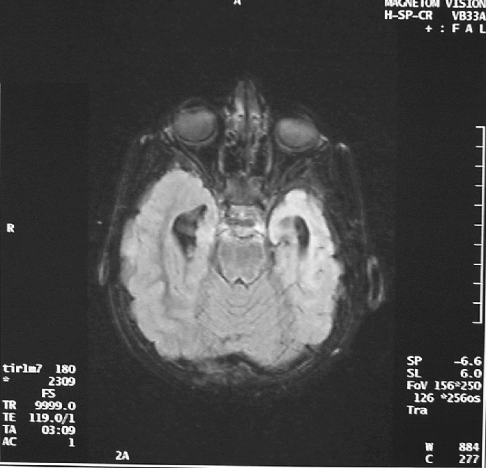
View fourth image(mr). Brain MR (Inversion Recovery Axial Image)
Full history/Diagnosis is available below
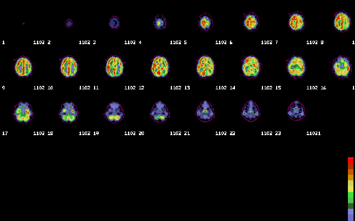
The patient was sent for an inter-ictal PET scan to evaluate for seizure focus for possible surgical resection. The patient was being monitored via EEG during the uptake phase of the PET FDG brain scan. During the uptake, the patient stated she was having a seizure. However, the EEG was unremarkable and the physicians thought the patient was continuing with her well-known habit of being manipulative in an attempt to get out of having to undergo the PET scan. The parents stated, however, that the child was exhibiting the stare characteristic of her seizure episodes and thought the patient was likely truly having a seizure.
However, if the patient has a seizure during the uptake phase of the PET study and if the seizure is of sufficient duration, the seizure focus may demonstrate increased FDG accumulation. In this case, several factors were present that make localization of the seizure focus easier. First, the patient's seizure occurred during the uptake phase. Second, the pateint's seizure was of sufficient duration such that the focus was able to take up a sufficient amount of radiopharmaceutical. Finally, the seizure focus did not generalize during the uptake phase thereby localizing uptake to a small focus corresponding to the site of origination of the seizure.
If the patient's seizure had not occurred duirng the uptake phase or had it not been of sufficient duration, there might not have been an opportunity for differential uptake of the FDG radiopharmaceutical to occur to delineate the seizure focus.
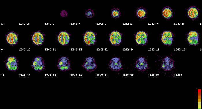
Second study is an inter-ictal PET scan obtained approximately five weeks later. This scan again demonstrates decreased activity in the left hemisphere globally with the more prominent decrease in the left parietooccipital region, all unchanged from the prior exam. However, the left temporal lobe is now hypometabolic. The patient did not report seizure activity during the uptake phase and thus the expected decrease in the left temporal lobe confirms this site as the originating focus for the seizure as decrease activity is expected in a seizure focus on an inter-ictal study, as described above.
Brain MR performed at St. Louis Children's Hospital reveals bilateral dysmorphic small hippocampi, total absence of the corpus callosum, grey-matter heterotopias, and a dysmorphic temporal lobe with dysmorphic features extending into the left parietal region.
2) Ictal PET is difficult to perform as the patient must have a seizure during the uptake phase and the seizure must be of sufficient duration to allow for enough FDG accumulation to demonstrate the focus.
3) Ictal PET will likely be less effective in demonstrating the origin of seizure activity if the seizure rapidly generalizes during the uptake phase.
4) Ictal PET in this case was a fortuitous event.
References and General Discussion of PET Brain (Nontumor) Imaging Studies (Anatomic field:Skull and Contents, Category:Normal, Technique, Congenital Anomaly)
Return to the Teaching File home page.