Case Author(s): J. Philip Moyers and Farrokh Dehdashti, M.D. , 9/23/95 . Rating: #D3, #Q4
Diagnosis: Epileptogenic focus Left temporal lobe
Mesial temporal sclerosis
Brief history:
Intractable partial complex
seizures since childhood.
Images:

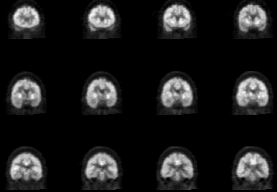
Coronal 18-FDG images of brain
View main image(pb) in a separate image viewer
View second image(mr).
Coronal MR image of brain, T2 weighted
View third image(mr).
Coronal MR image of brain, T2 weighted
Full history/Diagnosis is available below
Diagnosis: Epileptogenic focus Left temporal lobe
Mesial temporal sclerosis
Full history:
This is a 55-year old woman with
a history of complex partial seizures since childhood.
Her seizures have been intractable with medical
management. A recent MRI demonstrated small left
hippocampal formation and atrophic cerebellum. PET
imaging was requested as part of a presurgical
management plan.
Radiopharmaceutical:
F-18 fluorodeoxyglucose
Findings:
There is an area of hypometabolism
in the left temporal lobe and left thalamus. This
corresponds well to the findings of mesial temporal
sclerosis involving the medial aspect of the left
temporal lobe, hippocampal gyrus and amygdala. In
addition, generalized hypometabolism of the
cerebellum was seen, correlating with atrophic
changes seen on MRI. This is likely related to long-
term Dilantin therapy.
Discussion:
Complex partial seizures with a
temporal lobe focus are common among epileptic
patients. Imaging can be performed with CT, MRI, or
PET. MRI is superior to CT in identifying
morphologic abnormalities in patients with epilepsy.
Small areas of gliosis or small AVMs can be
overlooked with CT examination. Also, heterotopic
gray matter may also be missed by CT examination.
A cause of complex partial seizures is mesial temporal
sclerosis. In this pathologic entity, there is prominent
hippocampal cell loss with atrophy of the medial
aspect of the temporal lobe involved. In some patients
with partial seizures, MRI may be normal or
demonstrate a smaller temporal lobe without
increased signal intensity on the side of the lesion.
Therefore, interictal PET studies are helpful to
evaluate for interictal cerebral metabolism. Studies of
cerebral blood flow, metabolism, and neurotransmitter
activity are felt to be more promising than structural
imaging in the evaluation of patients with epilepsy
who have negative or unequivocal MRI. Clearly,
measurement of regional cerebral glucose metabolism
has proved the most useful in evaluation of patients
for seizure surgery. FDG-PET may avoid unnecessary
invasive tests. Most studies demonstrate interictal
hypometabolism in approximately 60-90% of patients
with complex partial seizures. The degree of
hypometabolism on the PET study is not related to
the percentage of neuronal loss and, the
hypometabolic regions are usually larger than the
area of pathologic abnormality. For example in the
patient with mesial temporal sclerosis where
hippocampal cell loss is a prominent feature,
hypometabolism involved the entire temporal lobe
rather than just the medial aspect in the region of the
hippocampal gyrus. Both qualitative and quantitative
analysis of PET studies can be used to lateralize the
abnormal temporal lobe for surgical resections,
although it is unclear which approach should be used.
In this patient, atrophy is demonstrated in the medial
aspect of the left temporal lobe consistent with the
diagnosis of mesial temporal sclerosis. Comparison
with the coronal PET images demonstrates
hypometabolism and decreased size of the left
temporal lobe when compared with the right temporal
lobe.
References:
1) Mazziotta JC, Gilman S. Clinical brain imaging:
principles and applications. Philadelphia, PA:
FA Davis Company, 1992.
2) Fisher RS, Frost JJ. Epilepsy. J Nucl Med
1991;32:651-659.
Followup:
MRI and monitored EEG suggest a
left temporal lobe seizure focus.
ACR Codes and Keywords:
References and General Discussion of PET Brain (Nontumor) Imaging Studies (Anatomic field:Skull and Contents, Category:Organ specific)
Search for similar cases.
Edit this case
Add comments about this case
Read comments about this case
Return to the Teaching File home page.
Case number: pb003
Copyright by Wash U MO

