Case Author(s): Gregg Schubach\Barry A. Siegel , 8/8/95 . Rating: #D4, #Q4
Diagnosis: Hemimegelencephaly of the brain
Brief history:
3-month old girl who has been
seizing approximately 50 times a day since birth.
Images:
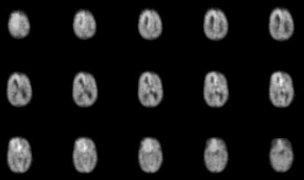
Axial PET Images
View main image(pb) in a separate image viewer
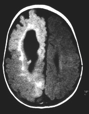
View second image(mr).
MRI through basal ganglia
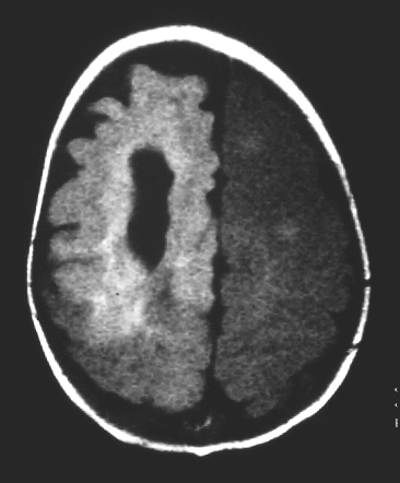
View third image(mr).
Axial MRI image at a higher level
View fourth image(mr).
Axial MRI near vertex
Full history/Diagnosis is available below
Diagnosis: Hemimegelencephaly of the brain
Full history:
3-month old girl referred for
assessment of right hemimegalencephaly, diagnosed
at the time of birth by MRI. The patient has
approximately 50 seizures daily, which are
characterized by deviation of the eyes to the left
with horizontal nystagmus and, on occasion,
deviation of the mouth to the left. Abnormal spike
EEG changes have been recorded from the
hemimegalencephalic and nonhemimegalencephalic
hemispheres.
Findings:
The PET images demonstrate
overall decreased activity in the brain compared
with the accumulation of FDG in the soft tissues of
the face. This overall decrease in cortical activity is
further confirmed when compared with the activity
in the region of the basal ganglia bilaterally. There
is a rind of relatively increased accumulation of
FDG in the region of the frontoparietal and inferior
frontal lobe on the right side. This corresponds with
the abnormally enlarged gyri seen on the MRI
study. The patient experienced a seizure within 30
minutes following the administration of FDG. The
hypermetabolism seen in the frontal aspect of the
hemimegalencephalic right hemisphere is
compatible with the increased FDG uptake during
the seizure or in close proximity to the recovery
phase of this or other seizures noted to have
occurred during the day of this study. The MRI,
which was performed approximately 2-1/2 weeks
prior to the PET study, demonstrates gyri to be abnormal
in both number and configuration. Post-
gadolinium images show contrast enhancement of
the frontal and parietal regions compared with
regions of normal parenchyma. The right cerebral
hemisphere is enlarged when compared with the left.
The frontal horn of the right lateral ventricle is
enlarged. Foci of gray matter heterotopia are noted
outlining the region of the left lateral ventricle.
Discussion:
Hemimegalencephaly is a
developmental brain malformation characterized by
congenital hypertrophy of one cerebral hemisphere
and ipsilateral ventriculomegaly with associated
epilepsy, hemianopsia, and varying degrees of
developmental delay. PET is a noninvasive method
used to study metabolism of the brain and has
proved to be useful in the understanding of many
neuropediatric syndromes and epilepsy. As
described by Rintahaka, et al., PET can provide
unique information that is useful in the management
of patients with hemimegalencephaly. There was
good correlation between CT/MRI and PET in
delineating areas of abnormality on the side of
hemimegalencephaly in their study of 8 children.
However, PET clearly revealed that many brain
regions, particularly on the
nonhemimegalencephalic side, which appear to be
normal structurally, were functionally abnormal.
Rintahaka P, Chugani
H, Messa C, Phelps M. Hemimegalencephaly:
evaluation with positron emission tomography.
Pediatric Neurology 1993;9:21-8.
Followup:
The patient subsequently
underwent partial right hemispherectomy (fronto-
parietal) two days following the PET study with
subsequent resolution of all seizure activity.
ACR Codes and Keywords:
References and General Discussion of PET Brain (Nontumor) Imaging Studies (Anatomic field:Skull and Contents, Category:Normal, Technique, Congenital Anomaly)
Search for similar cases.
Edit this case
Add comments about this case
Read comments about this case
Return to the Teaching File home page.
Case number: pb002
Copyright by Wash U MO