Case Author(s): Zhiyun Yang, M.D., Keith Fischer, M.D. , 06/14/04 . Rating: #D2, #Q3
Diagnosis: parathyroid adenoma
Brief history:
60 year-old woman with recurrent elevation of serum calcium and parathyroid hormone levels.
Images:
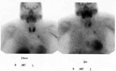
anterior 10 minute and 2 hour anterior planar images
View main image(pa) in a separate image viewer
View second image(pa).
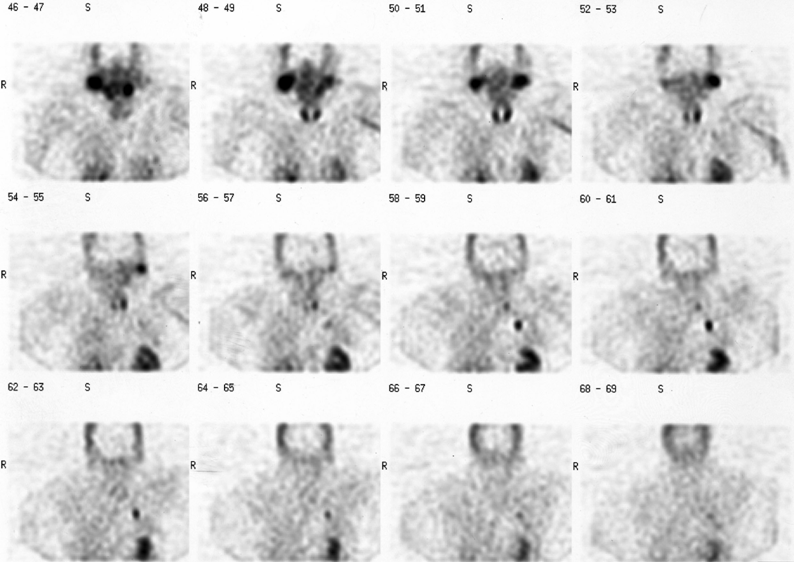
coronal SPECT images
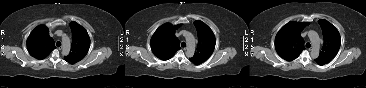
View third image(ct).
CT of the chest
Full history/Diagnosis is available below
Diagnosis: parathyroid adenoma
Full history:
60 year-old woman with a history of primary hyperthyroidism. She had resection of the right parathyroid gland performed appromimately 10 years ago. She now presents with recurrent elevation of serum calcium and parathyroid hormone levels.
Radiopharmaceutical:
19.9 mCi Tc-99m sestamibi i.v.
Findings:
After intravenous administration of Tc-99m sestamibi, planar images of the neck and mediastinum were obtained at approximately 10 minutes and 2 hours. Tomographic (SPECT) images of the neck and mediastinum were obtained immediately following the initial planar images.
There is an elongated focus of tracer accumulation noted posterior to the lower pole of the left thyroid lobe. Additionally, there is an intense focus of tracer accumulation in the left hemithorax. Correlation with chest radiographic examination performed the same day does not show a definite pulmonary nodule.
Discussion:
Increased tracer activity in the left hemithorax may represent activity in the left hilum or lung although no definite pulmonary lesion is identified on chest radiograph. Correlation with a computed tomography of the chest confirms that no lung lesion is present. This increased activity of tracer in the thorax has the following diferential diagnosis: 1.a mediastinal or ectopic parathyroid adenoma. 2. Neoplastic: such as: breast carcinoma, lung carcinoma, lymphoma, metastatic tumors. 3. Non--neoplastic, such as: actinomycosis, fibrosing alveolitis (diffuse), giant lymph node hyperplasia of the mediastinum.
Persistent focus of tracer accumulation posterior to the lower pole of the left thyroid lobe, is thought to represent a parathyroid adenoma.
Followup:
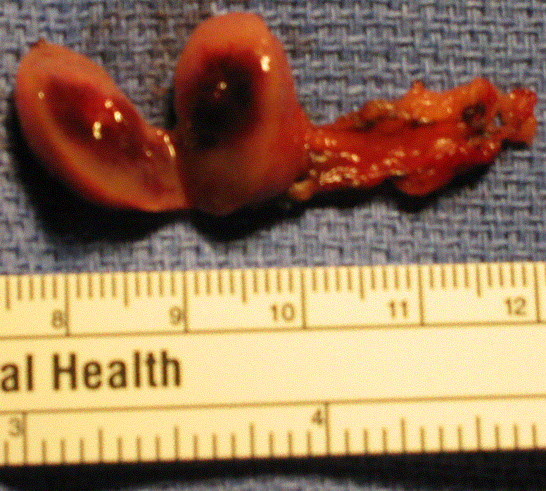
The first operation on 12-08-03 with neck re-exploration and subtotal (two and one-half gland)parathyroidectomy. 2.Transcervical left partial thymectomy.Pathologic report: parathyroid adenoma (0.8gm)from the left lower parathyroid position.
The second operation on 1-22-04 was a video-assisted excision of a mediastinal parathyroid adenoma. Pathologic report: parathyroid adenoma.
View followup image(mc).
Parathyroid adenoma
ACR Codes and Keywords:
- General ACR code: 23
- Face, Mastoids, and Neck:
2.363 "Adenoma"
References and General Discussion of Parathyroid Scintigraphy (Anatomic field:Face, Mastoids, and Neck, Category:Neoplasm, Neoplastic-like condition)
Search for similar cases.
Edit this case
Add comments about this case
Return to the Teaching File home page.
Case number: pa011
Copyright by Wash U MO