Case Author(s): Gabriel De Simon, M.D., Tom R. Miller, M.D., Ph.D. , 12/19/2000. . Rating: #D2, #Q4
Diagnosis: Parathyroid adenoma.
Brief history:
Asymptomatic female with elevated calcium and parathyroid hormone levels.
Images:
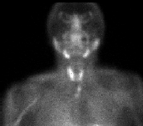
Ten-minute planar, anterior image of the neck and mediastinum.
View main image(pa) in a separate image viewer
View second image(pa).
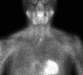
Two-hour delayed planar, anterior image of the neck and mediastinum.
View third image(pa).
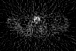
Transaxial 10-minute SPECT image at mid-neck level.
View fourth image(pa).
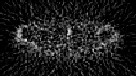
Transaxial 10-minute SPECT image at the sternoclavicular level.
Full history/Diagnosis is available below
Diagnosis: Parathyroid adenoma.
Full history:
Fifty-six year old, asymptomatic female with routine hematological examination demonstrating an elevated calcium level of 11 and parathyroid hormone value of 134.
Radiopharmaceutical:
21.6 mCi Technetium-99m sestamibi, i.v.
Findings:
INITIAL 10-MINUTE, ANTERIOR PLANAR IMAGE OF THE NECK:
Focus of increased radiopharmaceutical uptake located inferior to the caudad margin of the left thyroid lobe, separate from the thyroid gland.
Homogeneous uptake of radiotracer in a normal, non-enlarged thyroid gland.
DELAYED 2-HOUR, ANTERIOR PLANAR IMAGE OF THE NECK:
Persistence of activity in a small, rounded focus, located inferior to the caudad margin of the left thyroid lobe.
Normal thyroidal washout, with no significant residual activity.
INITIAL 10-MINUTE, TRANSAXIAL SPECT IMAGES AT THYROID AND STERNOCLAVICULAR LEVELS:
Focus of increased uptake just posterior to the left sternoclavicular joint, separate from the thyroid gland.
Discussion:
Technetium-99m sestamibi is a lipophilic, cationic radiopharmaceutical, used for both myocardial perfusion and tumor imaging. Sestamibi accumulates in both thyroid and parathyroid glands, in proportion to the regional blood flow, with peak activity four to six minutes after injection. The degree of sestamibi uptake by a parathyroid adenoma is independent of parathyroid hormone levels, but depends in part on gland size. Adenomas as small as 150 mg have been detected. The mechanism of sestamibi localization within the mitochondria and cytoplasm of an adenoma is in response to the electrical potentials generated across membrane bilayers. The large number of mitochondria in the cells of parathyroid adenomas may be responsible for the avid uptake and slow release of Tc-99 sestamibi in parathyroid adenomas compared to surrounding thyroid tissue. Knowledge of the embryological development of the parathyroid glands is essential in understanding location variations, a prerequisite for successful imaging. The inferior parathyroid glands, which originate from the third pharyngeal pouch, migrate caudally with the thymus, normally only as far as the inferior poles of the thyroid gland, but may descend with the thymus gland into the thorax. Approximately 90-95% of inferior parathyroid glands are located in the immediate vicinity of the lower pole of the thyroid gland, with ectopic sites including the superior thymic pole(39%), mediastinum(2%) and other miscellaneous sites. The position of the larger pair of superior parathyroid glands, which develop from the fourth pharyngeal pouch, is more constant, with 99% located behind the upper poles of the thyroid lobes. Aberrant superior PT glands may be found between the thyroid gland and esophagus, in the carotid sheath, behind the brachiocephalic vein or occasionally in the posterior mediastinum. The sensitivity of Tc-99m sestamibi in detection of parathyroid adenomas ranges from 88% to 100%, comparable to that of magnetic resonance imaging, and better than conventional imaging, including CT.
Followup:
At surgery, an aberrant left inferior parathyoid adenoma weighing 920 mg was found, embedded in the thyro-thymic ligament behind the posterior surface of the left sternocleidomastoid muscle, just above the level of the sternoclavicular joint, in the location demonstrated by the sestamibi scan. The adenoma was resected, with subsequent return of the patient's calcium and parathyroid hormone levels to normal.
Major teaching point(s):
1. Rationale behind single radionuclide technique in the imaging of parathyroid adenomas.
2. Embryological development of the parathyroid glands as a key factor to understanding the variations in location of the superior and inferior parathyroid glands, an important factor in successful imaging of parathyroid adenomas.
ACR Codes and Keywords:
References and General Discussion of Parathyroid Scintigraphy (Anatomic field:Face, Mastoids, and Neck, Category:Neoplasm, Neoplastic-like condition)
Search for similar cases.
Edit this case
Add comments about this case
Read comments about this case
Return to the Teaching File home page.
Case number: pa010
Copyright by Wash U MO