Case Author(s): Bart Rydzewski M.D., Ph.D. and Keith Fischer M.D. , 8/24/00 . Rating: #D2, #Q4
Diagnosis: Parathyroid adenoma
Brief history:
76 year old male with hypercalcemia
Images:
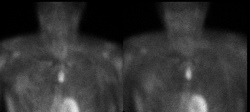
Anterior 10 min. and 2 hr delay images
View main image(pa) in a separate image viewer
View second image(pa).
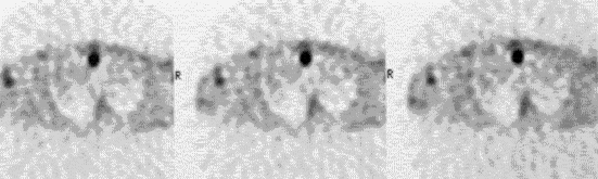
Axial SPECT images of the upper thorax
View third image(ct).
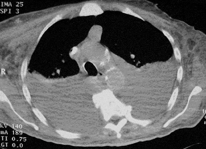
Axial CT images of the upper thorax
View fourth image(bs).
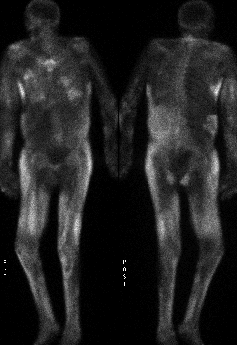
Whole body bone scintigraphy study.
Full history/Diagnosis is available below
Diagnosis: Parathyroid adenoma
Full history:
76 year old man with history of hypercalcemia, elevated serum parathyroid hormone level and end-stage renal failure who had a resection of 3-1/2 parathyroid glands recently yet has persistently elevated serum calcium and PTH. The study is requested to evaluate for residual or ectopic parathyroid tissue.
Radiopharmaceutical:
21.9 mCi Tc-99m sestamibi i.v.
Findings:
The planar parathyroid Tc-99m sestamibi scan (image #1) demonstrates a large oblong, vertically-oriented focus of increased activity in the midline of the upper chest on both early and delayed images. The abnormality is just above the level of the aortic arch. The position of the abnormal focus is confirmed on SPECT images of the neck and upper torso. This is consistent with a persistent mediastinal parathyroid adenoma. No other foci of increased activity are seen.
The computed tomography exam of the chest (image #3) demonstrates bilateral pleural effusions and an anterior mediastinal mass whose position and size correlates well with the scintigraphic abnormality described above. In addition, calcium deposition is evident in the subcutaneous tissues of the thorax (not shown).
Delayed bone scintigraphic images (image #4) demonstrate markedly abnormal distribution of the bone agent with diffuse uptake in the soft tissues. Lack of renal activity is due to the patient's end stage renal failure. There is activity linearly in the mid-abdomen, and diffusely in the thorax, presumably representing precipitation of calcium salts in the stomach and lungs respectively. Uptake of the radiopharmaceutical in the skeleton is very poor.
Discussion:
Tc-99m sestamibi has been widely used to evaluate patients for the presence of parathyroid adenomas. It has effectively replaced the previously popular Thallium-201/Tc99m-Pertechnetate method.
Sestamibi is a cationic complex found to localize in thyroid and parathyroid tissue. The kinetics of the compound are different for both tissues. After the initial uptake, there is no significant wash-out of sestamibi from parathyroid adenoma tissue which is presumably due to mitochondrial binding. In contrast, by 2 hours, there is marked reduction in sestamibi concentration within the thyroid tissue thus resulting in high parathyroid-to-thyroid uptake ratio of the radiopharmaceutical.
The standard examination consists of 10 minute delayed planar and SPECT images to demonstrate the anatomic relationship between the thyroid gland and parathyroid adenoma. This is followed by a 2 hour delayed planar image. The sensitivity of Tc-99m Sestamibi for detection of parathyroid adenoma has been quoted at 87% which compares with 71% for Thallium-201/Tc99m-Pertechnatate studies.
Extensive soft tissue calcifications seen in this patient are likely related to renal failure coupled with hypercalcemia. Deposition of calcium in the soft tissues occurring secondary to hypercalcemia is termed metastatic calcification. Metastatic calcification can result from any process with an elevated calcium-phosphate product. Entities such as renal failure, hyperparathyroidism, sarcoidosis, milk-alkali syndrome, etc. can lead to metastatic calcifications. These are often fine and diffuse throughout the soft tissues, especially heart, lungs, blood vessels, kidneys, brain, eyes, peri-articular tissues, and skin.
In severe cases, blood vessel deposits may lead to gangrene of the extremities. If the hyperphosphatemia is corrected, there is some reversal of the process. The risk increases when hyperphosphatemia is present for an extended period and the calcium (mg/dL)-phosphate (mg/dL) product exceeds 70.
Clinical Nuclear Medicine Ed. MN Maisey, KE Britton, BD Collier Chapman and Hall Medical 3rd edition 1998
Followup:
At surgery, a parathyroid adenoma was found at the location indicated both by the scan and CT.
ACR Codes and Keywords:
References and General Discussion of Parathyroid Scintigraphy (Anatomic field:Face, Mastoids, and Neck, Category:Neoplasm, Neoplastic-like condition)
Search for similar cases.
Edit this case
Add comments about this case
Return to the Teaching File home page.
Case number: pa009
Copyright by Wash U MO