Case Author(s): John Lahorra and Jerold Wallis , . Rating: #D2, #Q3
Diagnosis: Parathyroid adenoma
Brief history:
Elevated calcium
Images:
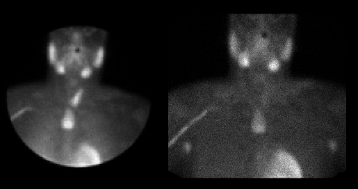
Initial and 2-hour delayed images
View main image(pa) in a separate image viewer
Full history/Diagnosis is available below
Diagnosis: Parathyroid adenoma
Full history:
34 yo with persistant hyperthyroidism s/p 2 resections with removal of 2 parathyroid glands and right thyroid lobectomy
Radiopharmaceutical:
Tc-99m Sestamibi
Findings:
Absent uptake is seen in the right thyroid lobe, consistent with prior resection. A large focus of increased uptake is seen in the superior mediastinum, which persists on delayed imaging.
Discussion:
Parathyroid adenomas can be found in the mediastinum. Imaging should include the chest (to the level of the myocardium), and just obtaining pinhole images of the thyroid region is not sufficient for the imaging study.
SPECT imaging or SPECT-CT would be helpful for localizing the depth of the adenoma, to aid in surgical planning. If separate CT is planned later, the level of the lesion can be marked on the skin after localizing it using a radioactive marker.
Followup:
CT showed a homogeneously enhancing 2.3 x 3.5 cm mass which was retrosternally located and extended superiorly to the posteroinferior aspect of the left lobe of the thyroid and inferiorly to just above the level of the aortic arch. This was surgically resected.
ACR Codes and Keywords:
References and General Discussion of Parathyroid Scintigraphy (Anatomic field:Face, Mastoids, and Neck, Category:Neoplasm, Neoplastic-like condition)
Search for similar cases.
Edit this case
Add comments about this case
Return to the Teaching File home page.
Case number: pa002
Copyright by Wash U MO