Case Author(s): J. Wallis , 8/4/94 . Rating: #D3, #Q3
Diagnosis: Mediastinal parathyroid adenoma
Brief history:
Patient with elevated calcium levels.
Images:
View main image(pa) in a separate image viewer
Full history/Diagnosis is available below
Diagnosis: Mediastinal parathyroid adenoma
Full history:
The patient has had neck exploration for a suspected
parathyroid adenoma, with removal of one normal size
parathyroid gland. He has persistantly elevated
calcium levels.
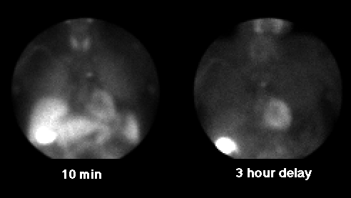
Findings:
The patient was imaged with Tc-99m Sestamibi. Initial
images show normal uptake in the thyroid gland, heart,
liver, and bowel; in addition there is
a focus of uptake in the mediastinum. Delayed images
show normal washout of tracer from the thyroid gland,
without a persistant focus in the thyroid bed to suggest
a parathyroid adenoma. However, the mediastinal
focus is again seen.
Discussion:
The findings suggest an ectopic parathyroid adenoma in
the mediastinum.
Followup:
A marker was placed over the site of mediastinal uptake,
and subsequent CT imaging confirmed a 1 cm enhancing
nodule in the anterior aspect of the superior mediastinum
suggesting a parathyroid adenoma. The parathyroid
adenoma was resected surgically.
Major teaching point(s):
1) Images for parathyroid adenomas should include views
of the chest and mediastinum as well as the neck.
2) Sestamibi is a useful agent for parathyroid imaging.
When imaging non-ectopic parathyroid adenomas, the slower
washout of tracer
from parathyroid tissue compared to normal thyroid tissue
aids in identification of adenomas.
ACR Codes and Keywords:
References and General Discussion of Parathyroid Scintigraphy (Anatomic field:Lung, Mediastinum, and Pleura, Category:Neoplasm, Neoplastic-like condition)
Search for similar cases.
Edit this case
Add comments about this case
Read comments about this case
Return to the Teaching File home page.
Case number: pa001
Copyright by Wash U MO