Case Author(s): Xia Wang, M.D., Quan Vu, M.D., Keith Fischer, M.D. , 06/25/06 . Rating: #D2, #Q4
Diagnosis: Terminal Ileum Carcinoid Tumor.
Brief history:
A 50-year-old male with terminal ileum mass.
Images:

Octreotide Scintigraphy, Coronal Plannar Image.
View main image(ot) in a separate image viewer
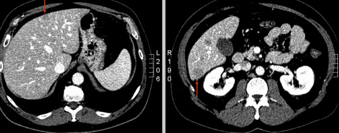
View second image(ct).
Axial CT Images of Lower Abdomen.
View third image(ct).
Axial CT Images of Liver.
Full history/Diagnosis is available below
Diagnosis: Terminal Ileum Carcinoid Tumor.
Full history:
This is a 50-year-old male with diagnosis of carcinoid tumor in the terminal ileum and numerous hepatic lesions per an abdominal pelvic CT.
Radiopharmaceutical:
5.7 mCi In-111 Pentetreotide i.v.
Findings:
There are innumerable foci of intense In-111 activity scattered throughout the liver, likely corresponding to the hypoattenuating liver lesions seen on the patient's recent CT exam, likely representing metastases. A focus of intense uptake is also seen in the right lower quadrant in the expected position of the terminal ileum, likely representing the patient's primary lesion.
CT findings: In the right lower quadrant, there is an ill defined mesenteric mass/desmoplastic reaction which measures approximately 1.7 x 1.4 cm. Adjacent to this region is mild dilation of the terminal ileum as well as fatty replacement and thickening of the colonic and cecal wall. There are numerous round, well defined lesions involving all segments of the liver.
Discussion:
Neuroendocrine tumors contain somatostatin receptors. This feature allows for the localization of primary tumors and tumor metastases by scintigraphy with the radiolabeled somatostatin analog octreotide. It can be used to detect certain types of cancer arising from the neuro-endocrine system, such as non-secreting pancreatic islet cell tumors, gastrinomas, paragangliomas, pheochromocytomas, neuroblastomas, carcinoids, and Merkel cell carcinomas.
Whole-body images are obtained 4 hours and 24 hours after injection of In-111 pentetreotide. Tomographic (SPECT) images can be obtained at 4 hours for the abdomen and pelvis and 24 hours for the chest. Repeat SPECT images of the pelvis and abdomen can be performed at 24 hours but administration of cathartics the evening before is advised.
Followup:
Patient underwent ternimal ileum and ileocolic resection. Surgical pathology confirmed a terminal ileum carcinoid tumor.
Major teaching point(s):
In-111 Pentetreotide attaches to somatostatin receptor subtypes that are plentiful in carcinoid tumors so these tumors are seen with a high sensitivity with this agent.
ACR Codes and Keywords:
References and General Discussion of Octreotide Scintigraphy (Anatomic field:Gasterointestinal System, Category:Neoplasm, Neoplastic-like condition)
Search for similar cases.
Edit this case
Add comments about this case
Return to the Teaching File home page.
Case number: ot010
Copyright by Wash U MO