Case Author(s): Zhiyun Yang, M.D. and Jerold Wallis, M.D. , 04/01/05 . Rating: #D3, #Q3
Diagnosis: Metastatic neuroendocrine carcinoma
Brief history:
65-year-old male presents for assessment of tumor disease.
Images:
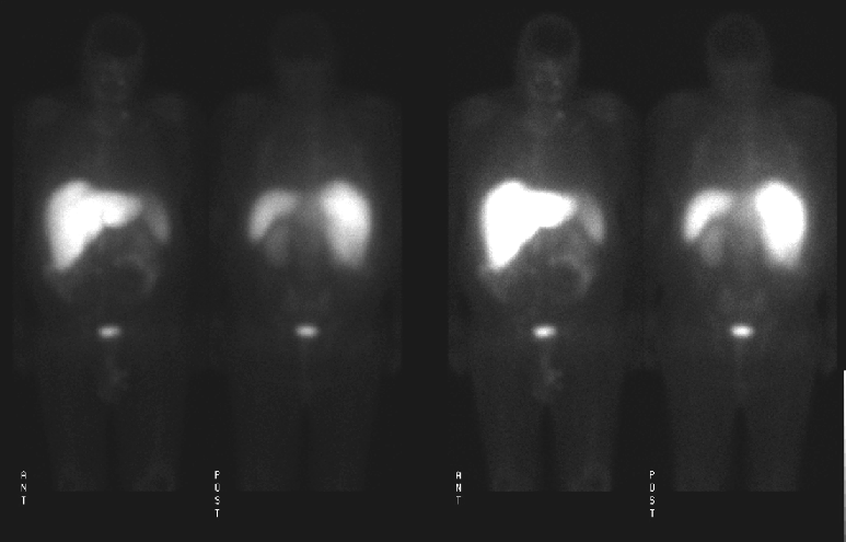
4-hour whole body scan
View main image(ot) in a separate image viewer
View second image(ot).
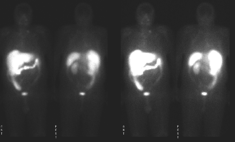
24 hours whole body scan
View third image(ot).
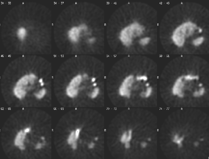
24 SPECT images
View fourth image(ct).
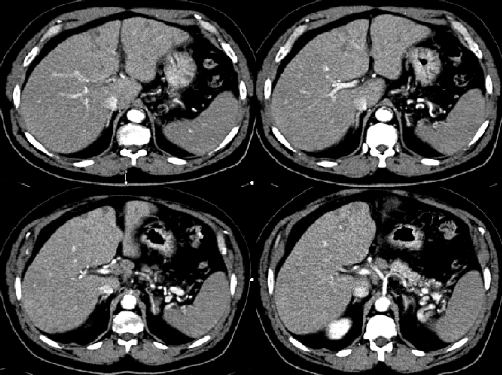
Selected CT axial images
Full history/Diagnosis is available below
Diagnosis: Metastatic neuroendocrine carcinoma
Full history:
65-year-old male who has poorly differentiated neuroendocrine carcinoma of the liver by biopsy. This examination was obtained to evaluate for additional sites of metastatic disease and for assessment of possible primary site of disease.
Radiopharmaceutical:
5.71 mCi In-111 pentetreotide i.v.
Findings:
Whole-body images were obtained 4 hours and 24 hours after injection of In-111 pentetreotide. Tomographic (SPECT) images of the abdomen were obtained at 4 (not shown) and 24 hours. There is expected In-111 pentetreotide activity in the spleen, kidneys. The relative activity in the liver, as compared to the kidneys, is markedly increased diffusely. Diffuse, markedly increased liver activity suggestive of diffuse liver infiltration. Mild activity in the aortocaval and left supraclavicular regions is consistent with lymph node involvement
The CT study demonstrated that several non-enhancing ill-defined infiltrative lesions seen throughout the liver, predominantly in the left lobe. Lymphadenopathy in the peripancreatic and retroperitoneal regions is also present.
Discussion:
Pentetreotide is a somatostatin analogue which normally accumulates in the thyroid and salivary glands, spleen, liver, and kidneys, with excretion into urinary bladder and bowel(mainly on 24 hour images). Kidneys and spleen retain the most activity and kidneys are seen even on delayed views. On delayed 24 hour image, regional uptake should be about threefold lower in the liver than in the spleen or kidneys. However, in this case, the activity of the liver is markedly increased compared with the activity in the spleen and kidney.
References:
1. Leners N, Fiasse R, et al. Indium-111-pentetreotide uptake in endocrine tumors and lymphoma. J. Nucl. Med. 1996; 37: 6916-922.
2. Mettler FA. Essentials of Nuclear Medicine Imaging. Fourth Edition. W.B. Saunders Company. 1998. 381.
Followup:
Core biopsy of the liver demonstrated that sections of liver show extensive involvement by metastatic well-differentiated neuroendocrine carcinoma. The tumor cells are uniform bland, round nuclei and are arranged in nests and ribbons. Occasional rosettes are seen. The primary differential diagnosis includes a pancreatic endocrine tumor or carcinoid tumor
Major teaching point(s):
Knowing normal biodistribution of In-111 pentetreotide is key in this case.
ACR Codes and Keywords:
References and General Discussion of Octreotide Scintigraphy (Anatomic field:Gasterointestinal System, Category:Neoplasm, Neoplastic-like condition)
Search for similar cases.
Edit this case
Add comments about this case
Return to the Teaching File home page.
Case number: ot009
Copyright by Wash U MO