Case Author(s): Akash Sharma, M.D. and Keith C. Fischer, MD , . Rating: #D2, #Q3
Diagnosis: Diffuse peritoneal metastases from Merkel cell tumor
Brief history:
76-year-old male presents for follow-up evaluation for metastatic disease with new abnormalities on a recent CT.
Images:

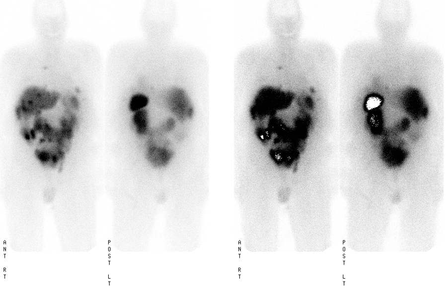
Whole body 24 hour planar images from an Octreotide study
View main image(ot) in a separate image viewer
View second image(ct).
Selected images from a recent abdomen CT performed prior to the Octreotide study.
View third image(ot).
Selected 24-hour axial SPECT images of the abdomen
View fourth image(ot).
Selected 24-hour coronal SPECT images of the abdomen
Full history/Diagnosis is available below
Diagnosis: Diffuse peritoneal metastases from Merkel cell tumor
Full history:
76-year-old gentleman with known metastatic neuroendocrine tumor, specifically Merkel cell tumor of the buttock, that has now involved the left adrenal and multiple sites in the abdomen. The patient has had radiation treatment to the retroperitoneal disease. The patient presents for follow-up evaluation for metastatic disease with new abnormalities on a recent CT. The abdomen CT was performed at Barnes-Jewish Hospital on 09/05/2003.
Radiopharmaceutical:
6.38 mCi In-111 pentetreotide i.v.
Findings:
ABDOMEN CT:
1. Marked expansion of left renal mass now into the pelvis with partial mechanical obstruction of the distal rectosigmoid colon. The mass now appears to encase both kidneys with hydronephrosis on the right.
2. Interval placement of a left kidney stent.
3. Interval development of retrocrural lymphadenopathy, perihepatic and perisplenic ascites with peritoneal implants in the right upper quadrant.
OCTREOTIDE SCINTIGRAPHY:
1. Single focus of activity in the right hilum/mediastinal region, multiple foci projecting over the liver which on SPECT images are actually on the surface of the liver. Another focus of activity is seen in the left upper quadrant projecting anteriorly and superiorly to the spleen. Mild to moderate bowel activity was seen on the initial 4 hour images. The patient has a known horseshoe kidney with radionuclide activity seen in the renal parenchyma.
2. There is relatively low level uptake of Octreotide in the primary tumor in the retroperitoneal region as well as in the left adrenal region. This may be secondary to patient's previous radiation treatment.
3. Prominent bowel activity in the left hemipelvis is likely a sequela of obstruction from the patient's known large mass.
4. Additional small foci of increased activity specifically in the right side of the mediastinum and at the left lung base. These are evidence for supradiaphragmatic metastases from the patient's known Merkel cell tumor.
Discussion:
Octreotide scans are useful in many neuroendocrine tumors as many neuroendocrine tumors have somatostatin receptors. Injection of the somatostatin receptor imaging agent that is coupled with a radioisotope will selectively go to neuroendocrine tumors. This is an important study for localizing neuroendocrine tumors that are not visible on other modalities such as CT and MRI imaging. Carcinoid tumors and gastrinomas are two tumors readily seen with Octreotide.
References:
Kvols LK. Somatostatin-receptor imaging of human malignancies: a new era in the localization, staging, and treatment of tumors.
Gastroenterology. 1993 Dec;105(6):1909-11.
Followup:
The patient had progressive spread of his cancer and ultimately had oliguric acute renal failure with respiratory failure. He expired during this hospital admission.
Major teaching point(s):
As evident by the thoracic activity in this case which was not commented on by CT, octreotide scintigraphy is more sensitive than conventional CT for detecting small deposits of metastatic disease of neuroendocrine origin. While ultimately inconsequential in this particular case, the increased sensitivity may be helpful in diagnosis and followup of small tumors.
ACR Codes and Keywords:
References and General Discussion of Octreotide Scintigraphy (Anatomic field:Gasterointestinal System, Category:Neoplasm, Neoplastic-like condition)
Search for similar cases.
Edit this case
Add comments about this case
Return to the Teaching File home page.
Case number: ot008
Copyright by Wash U MO

