Case Author(s): Jerold Wallis , 5/31/94 . Rating: #D3, #Q3
Diagnosis: VIPoma metastatic to liver
Brief history:
Patient with history of a VIPoma. Assess for residual disease
Images:
View main image(ot) in a separate image viewer
Full history/Diagnosis is available below
Diagnosis: VIPoma metastatic to liver
Full history:
The patient is status post resection of a pancreatic
VIPoma, and subsequent resection of a solitary liver
metastasis one year ago. The patient now has recurrent
symptoms. MR examination of the abdomen
did not reveal any tumor foci.
Findings:
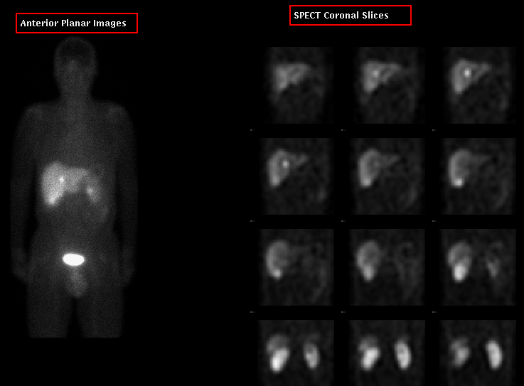
There are two abnormal areas of increased radiopharmaceutical uptake
in the liver, one near the junction of the right and left lobes, and the other
in the posterior segment of the right liver lobe near the inferior liver
edge. These almost certainly represent metastatic disease from the
patient's prior VIP secreting tumor.
Discussion:
Octreotide imaging is useful for detecting a variety
of neuro-endocrine tumors which contain somatostatin
receptors, including VIPomas,
insulinomas, gastrinomas and pheochromocytomas.
Followup:
An angiographic CT examination confirmed the two liver lesions, and also
demonstrated several additional small lesions within
the liver.
Major teaching point(s):
Uptake at the inferior liver margin on the planar images
could have been due to (normal) hepatobiliary excretion of
tracer. SPECT is useful to localize this activity further;
the relatively posterior location suggests a tumor focus
within the liver.
ACR Codes and Keywords:
References and General Discussion of Octreotide Scintigraphy (Anatomic field:Gasterointestinal System, Category:Neoplasm, Neoplastic-like condition)
Search for similar cases.
Edit this case
Add comments about this case
Read comments about this case
Return to the Teaching File home page.
Case number: ot001
Copyright by Wash U MO