Return to Case List with Diagnoses or Case List as Unknowns

PYOMYOSITIS
Authored By: Farrokh Dehdashti and Xiaoni Hong.
Patient: 55 year old female
History: This is a 55-year-old woman who was in her usual state of good health until approximately 1-1/2 weeks ago when she developed fever, back pain, and symptoms of malaise and an upper respiratory infection.
Image Size:[small][as-submitted]

Fig. 1
Coronal PET images of the lower extremities reveal intense FDG uptake in the knee joints and various muscles in a pattern most compatible with infection.

Fig. 2
PET/CT images of the lower extremities reveal large necrotic-appearing masses within each of the gastrocnemius muscles. Each is about 5 cm in diameter and measures 15 Hounsfield units, surrounded by a rim of marked FDG avidity. These are both most compatible with gastrocnemius abscesses or focal muscle liquefaction. Further FDG uptake is seen along the course of the right anterior tibialis muscle. There is a focus of marked FDG avidity in the right mid-foot and at the lateral aspect of the left forefoot.The right gluteal musculature is grossly enlarged with indistinct margins, also demonstrating moderate to marked FDG avidity. There is moderate uptake of FDG within the right hamstring muscle group. Marked FDG uptake is observed in a synovial distribution surrounding both knees.

Fig. 3
Axial MRI T1 constrast of Lower extrimities reveal bilateral, moderate to large, fluid collections in both gastrocnemius muscles with smooth peripheral rim enhancement after contrast administration.
Image Size:[small][as-submitted]

Findings:

FDG-PET/CT findings: a large necrotic-appearing mass within the gastrocnemius muscles bilaterally. Each is about 5 cm in diameter and measures 15 Hounsfield units, surrounded by a rim of marked FDG activity. These findings are most compatible with intramuscular abscesses or focal muscle liquefaction. Further FDG uptake is seen along the course of the right anterior tibialis muscle. There is a focus of marked FDG avidity in the right mid-foot and at the lateral aspect of the left forefoot.The right gluteal musculature is grossly enlarged with indistinct margins, also demonstrating moderate to marked FDG uptake. The medial aspect of the left psoas muscle demonstrates increased FDG uptake along its entire course. There is moderate uptake of FDG within the right hamstring muscle group. Marked FDG uptake is observed in a synovial distribution surrounding both knees.

MRI of Lower extrimities: bilateral, moderate to large, fluid collections in both gastrocnemius muscles with smooth peripheral rim enhancement after contrast administration. There is mild enhancing synovitis and moderate knee effusions bilaterally.

A large, multiloculated fluid collection within the left vastus intermedius demonstrates thick enhancing septa on the post contrast sequences. This pattern most likely represents myositis with confluent microabscesses. A similar pattern is seen in the right vastus intermedius but to a somewhat less extent.


Diagnosis: Pyomyositis
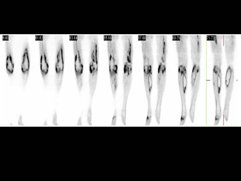
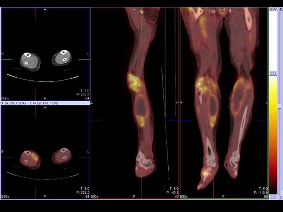
General Discussion:

Radiopharmaceutical: 13.2 mCi F-18 Fluorodeoxyglucose i.v.

Long History history: This is a 55-year-old woman who was in her usual state of good health until approximately 1-1/2 weeks ago when she developed fever, back pain, and symptoms of malaise and an upper respiratory infection. She presented to her primary physician who prescribed non-steroidal anti-inflammatory medications. She subsequently developed severe joint pain of the lower extremities and bilateral leg swelling. She is on no other medications and reports no history of malignancy. The patient was admitted for further evaluation. A chest CT was acquired that demonstrated numerous subcentimeter indeterminate pulmonary nodules in both lungs that were concerning for pulmonary metastases of an unknown primary.


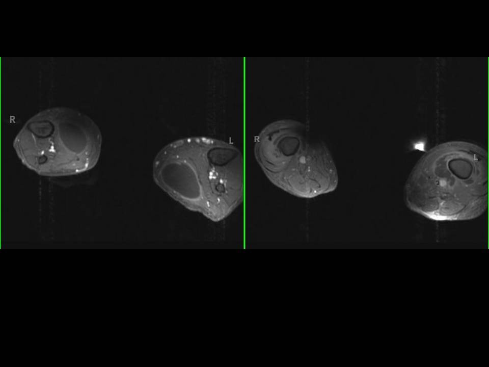
Discussion: Pyomyositis, a purulent infection of skeletal muscle that is usually caused by Staphylococcus aureus.Once considered a tropical disease, it is now seen in temperate climates as well, particularly with the emergence of HIV infection. In addition to HIV, other viruses, bacteria (including mycobacteria), fungi, and parasites can cause myositis. Single or multiple muscle groups in the limbs can be involved. MRI is helpful in differentiating pyomyositis from osteomyelitis. FDG-PET is non-specific and complementay to CT or MRI for diagnosis pyomyositis.

Follow up:  cytology of right calf muscle demonstrated cellular debris and acute inflammation. Blood culture was positive for STAPHYLOCOCCUS AUREUS. he underwent I&D of the bilateral calf muscles, collections as well as arthroplasty and I&D of her
bilateral septic knee joints. Evaluation for endocarditis which was the presumed origin given her multiple distal abscesses revealed no echocardiographic evidence of vegetation on her heart valves by transthoracic echocardiogram.  Cardiac Echo demonstrated right to left shunt. Patient was treated Oxacillin for presumed bacterial endocarditis and discharge home.

References: 1. Bleeker-Rovers CP, Vos FJ, Corstens FH, Oyen WJ. Imaging of infectious diseases using [18F] fluorodeoxyglucose PET. Q J Nucl Med Mol Imaging. 2008 Mar;52(1):17-29
Comments:
No comments posted.
Additional Details:

Case Number: 206898Owner(s): Farrokh Dehdashti and Xiaoni HongLast Updated: 12-07-2011
Anatomy: Skeletal System   Pathology: Infection
Modality: PETAccess Level: Readable by all users, writable by NucMed Certifiers
Keywords: ptnmACR: 40000.61400

Case has been viewed 53 times.
Certified by Farrokh Dehdashti on 07-07-2008

The reader is fully responsible for confirming the accuracy of this content.
Text and images may be copyrighted by the case author or institution.