Return to Case List with Diagnoses or Case List as Unknowns

CALYCEAL RUPTURE DUE TO ACUTE URETERIC OBSTRUCTION
Authored By: Keith Fischer and Dhanashree Rajderkar.
Patient: 58 year old female
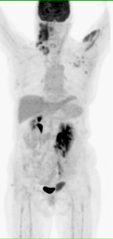
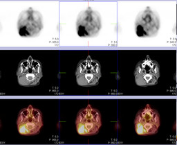
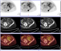
History: 50-year-old woman with acute myeloid leukemia, treated with stem cell transplant. A recent biopsy of a right neck mass demonstrated myeloid sarcoma.PET-CT was requested to stage the disease.
Image Size:[small][as-submitted]

Multimedia: 326971_1_submitted.avi

Fig. 2

Fig. 3

Fig. 4

Fig. 5

Fig. 6

Fig. 7

Fig. 8

Fig. 9

Fig. 10

Fig. 11
Image Size:[small][as-submitted]

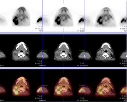
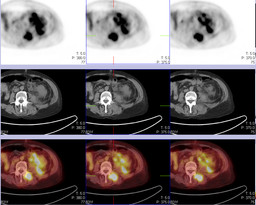
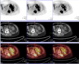
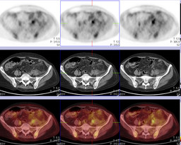
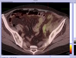
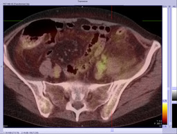
Findings:

 FDG-PET: There is a large soft tissue mass in the posterior right neck, representing a conglomerate of
enlarged posterior cervical lymph nodes measuring 6.8 x 4.5 cm collectively.  There is intense FDG uptake within this mass. (Maximum SUV 8.2) In addition, there are hypermetabolic metastatic deposits within the left biceps brachialis and pectoralis muscles, bilateral supraclavicular lymph nodes, left paraspinal muscles at L4-L5, and nodes in the left groin (maximum SUV of 6.4).

There are also foci of increased FDG uptake within the lower abdomen and pelvis along the course of the left ureter, most consistent with iliac chain lymph node deposits. The proximal left ureter is dilated, and there is left hydronephrosis. In addition, inflammatory stranding and free fluid surround the left kidney; there is extrarenal extravasation of FDG into the retroperitoneum, from forniceal rupture.

Impression: Extensive metabolically active soft tissue deposits representing myeloid sarcoma within the neck, left upper extremity, abdominal wall, lumbar paraspinal muscles, and left iliac chain lymph nodes, the latter of which caused left-sided hydroureteronephrosis and forniceal rupture.


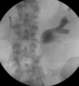
Left sided retrograde pyelogram followed by stent placement was performed.


DDx: 1.Extensive metastases from known myeloid sarcoma. Acute left ureteric obstruction with calyceal rupture.
Diagnosis: Acute calyceal rupture due to acute left distal ureteric obstruction by enlarged metastatic lymph nodes.
General Discussion:

Rupture of the urinary collecting system with peripelvic extravasation of urine may produce either an encapsulated colection of urine called urinoma which is often asymptomatic or urinary ascites and an acute abdomen.

Rupture of the fornix of a calyx leads to backflow of urine and contrast material into the renal sinus. Once in the renal sinus, the extravasate may enter the lymphatics (pyelolymphatic backflow) or veins (pyelovenous backflow) or it may course around the pelvis into the retroperitoneum (peripelvic extravasation).

Forniceal rupture occurs almost equally on both the right and the left sides. The male : female ratio is 3/1.

Most cases of extravasation have been associated with ureteral obstruction by calculi.

Other causes include trauma and invasive urologic procedures, posterior uretheral valves in neonates, pregnancy, and benign prostatic hypertrophy. Tumors have included primary ureteral neoplasms, carcinoma of the bladder and cervix, and metastatic disease from the stomach, breast, and colon. Other causes are pelvic-ureteric junction (PUJ) obstruction, vascular extrinsic compression, iatrogenic and i.v. fluid administration.

The most common etiology of renal forniceal rupture is obstruction caused by distal ureteric stones followed by malignant extrinsic ureteric compression. 

Extravasated urine incites a low-grade inflammatory reaction followed by an avascular deposition of collagen and fibrous tissue accounting for the urinoma formation. It has been postulated that all cases of hydronephrosis have mild pyelosinus extravasation and that the urine causes a local fibrotic response which prevents further disruption of the fornical area. This fact may account for the relatively few cases of peripelvic extravasation in adults with chronic urinary tract obstruction. However peripelvic urinary granulomas, retroperitoneal fibrosis, perinephric abscess, urinomas  and electrolyte disturbances may also occur. 

Placement of an indwelling ureteral stent or diversion via a percutaneous catheter can relieve obstruction from tumor compression. Nephrectomy may be indicated in cases with substantial loss of renal function and with a normal contralateral kidney. When the urinary tract obstruction is relieved there is usually resolution of radiographically evident backflow.

References:

1.Gennaro Cormio, M.D., Luigi Cormio, M.D., Ph.D.,*,1 Giuseppe Di Gesu’, M.D., Giuseppe Loverro, M.D.,and Luigi Selvaggi, M.D. CASE REPORT: Calyceal Rupture and Perirenal Urinoma as a Presenting Sign of Recurrent Ovarian Cancer; Gynecologic Oncology 83, 415–417 (2001)

2.Phornthep Ruchdaphornkul, Kamol Panumatrassamee; Clinical report:Transitional cell carcinoma of ureter presenting acute ureteric colic and ruptured caliceal fornix with extravasation of urine; Asian Biomedicine Vol. 4 No. 6 December 2010; 935-938.

3.Eric J. Heffernan and Stephen J. Skehan;INTERESTING IMAGE: Calyceal Rupture Secondary to Ureteric Obstruction by recurrent Colorectal Carcinoma; Clin Nucl Med 2007;32: 199–200.

4.http://emedicine.medscape.com/article/382530-overview#a23

5.Boris Gershman, Naveen Kulkarni, Dushyant V. Sahani and Brian H. Eisner; Causes of renal forniceal rupture; 2011 BJU INT ERNATIONAL 

Comments:
No comments posted.
Additional Details:

Case Number: 326971Owner(s): Keith Fischer and Dhanashree RajderkarLast Updated: 01-30-2013
Anatomy: Genitourinary (GU)   Pathology: Other
Modality: GU, PETAccess Level: Readable and writable by Nuclear Medicine only
Keywords: calyceal rupture

Case has been viewed 20 times.
Certified by Keith Fischer on 01-30-2013

The reader is fully responsible for confirming the accuracy of this content.
Text and images may be copyrighted by the case author or institution.