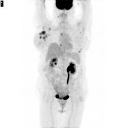
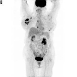
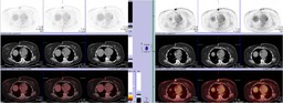
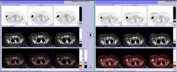
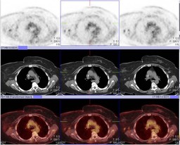
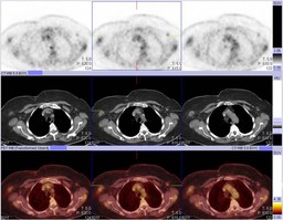
Return to Case List with Diagnoses or Case List as Unknowns

PET ESTRADIOL CHALLENGE TEST
Authored By: Farrokh Dehdashti and Archana Kantawala.
History: 66-year-old female with stage IV left breast cancer.ER positive, PR positive post 12 cycles of chemotherapy. This study was performed to see if a patient’s breast cancer has active functional estrogen receptors and may benefit from hormonal therapy
Image Size:[small][as-submitted]

Fig. 1
Day1 PET

Fig. 2
Day 1 PET coronals

Fig. 3
Day 2 PET after estradiol challenge

Fig. 4
Day2 coronals

Fig. 5
Day 1 on the left, Day 2 on the right

Fig. 6
Day 1 on the left. Day 2 on the right

Fig. 7
PET 3 after completion of chemotherapy

Fig. 8
PET 3 coronals

Fig. 9
PET 3 axials

Fig. 10
PET 3 axials
Image Size:[small][as-submitted]

Findings:

PET day 1 - FDG avid lesion in the lower outer quadrant of the left breast. Nodular skin thickening in both breasts, left greater than right with bulky axillary (right greater than left), mediastinal, supraclavicular, and right posterior triangle lymphadenopathy.There is a second FDG avid lesion in the central right breast.

PET day 2 -Overall, majority of previously seen lesions (PET/CT day1) show only slight increase uptake compared with the prior study. There is an overall of only 9% increased in FDG uptake in these lesions after estradiol therapy. This is NOT indicative of metabolic flare after estradiol therapy.

The lesions to be analyzed are selected on the first day PET study (typically up to 6 most intense lesions). The SUVmax of the lesion(s) is measured on both PET studies. The average SUVmax of the lesions on each day will be calculated and the percent change in FDG uptake will be determined.

≥ 12% increase in FDG uptake on the second day PET in comparison to the first day PET is indicative of "metabolic flare" and thus, the likelihood of response to hormonal therapy

 PET 3 -  Interval resolution of metaboilic activity suggesting response to chemotherapy.

Diagnosis:

 This is NOT indicative of metabolic flare after estradiol therapy.

Dehdashti et al. reported that an increased in FDG uptake greater than 12% after estradiol challenge is indicative of response to hormonal therapy (Breast Cancer Res Treat. 2009;113:509-17).

General Discussion:

 In light of the PET estradiol challenge test, the treatment plan was changed to chemotherapy. The patient underwent a PET study(PET 3) after completion of chemotherapy which showed marked response to therapy.

Most breast cancers are estrogen receptor (ER)-positive (ER+) tumors and therefore potentially hormone responsive. Despite this, chemotherapy often is chosen over hormonal therapy in the initial management of advanced breast cancer, at least in part because many physicians believe that chemotherapy is more likely to produce tumor shrinkage and to produce it more rapidly than hormonal agents. Even in elderly women, who tend to have a more indolent disease process, it has been reported that hormonal therapy is underused.In advanced ER+ breast cancer, the likelihood of response to hormonal therapy is equivalent to the likelihood of response to chemotherapy.

Hormonal therapies are consistently underused in the management of breast cancer, although the majority of patients have ER+ disease.The presence of ER alone predicts clinical benefit in 55% to 60% of patients. If both ER and PR are present, the likelihood of benefit increases only slightly, to 60% to 70%.The presence of a hormone receptor does not indicate that the receptor is functional and essential to the growth of the cancer cell, of course, nor does it imply that interference with receptor function will result in tumor-cell kill. None of the available serologies, prognostic factors, or radiologic studies can accurately predict for clinical benefit from hormonal therapy.

Hormone flare reactions seem to be more predictive of hormone responsiveness than the results of receptor assays, because flare indicates that the receptors are both present and functional. The flare reaction typically occurs in postmenopausal women with ER+ breast cancer and osseous metastases. Within 7 to 10 days of initiation of hormonal therapy, patients develop increased pain at metastatic sites, often accompanied by increased serum calcium, alkaline phosphatase, or tumor markers.Serial skeletal scintigraphy may show increased activity at sites of known metastasis or may identify areas not appreciated on pretreatment scintigrams. Although this clinical presentation often is indistinguishable from disease progression, true hormone-induced flare reactions herald subsequent response to treatment in 75% to 90% of patients. Nevertheless, clinical flare is of limited use in the prediction of response because it is uncommon (observed in less than 5% of patients), although it may occur at a subclinical level in more patients.

In patients with functional ER, metabolic flare can be seen following treatment with tamoxifen or after short treatment with estradiol.

References:

Journal of Clinical Oncology, Vol 19, Issue 11 (June), 2001: 2797-2803 © 2001 American Society for Clinical Oncology


Breast Cancer Res Treat. 2009 Feb;113(3):509-17.


JAMA. 2009 Aug 19;302(7):774-80

Comments:
No comments posted.
Additional Details:

Case Number: 324079Owner(s): Farrokh Dehdashti and Archana KantawalaLast Updated: 12-08-2011
Anatomy: Other   Pathology: Other
Access Level: Readable and writable by Nuclear Medicine only

Case has been viewed 26 times.
Certified by Farrokh Dehdashti on 12-08-2011

The reader is fully responsible for confirming the accuracy of this content.
Text and images may be copyrighted by the case author or institution.