Return to Case List with Diagnoses or Case List as Unknowns

METASTATIC MALIGNANT MELANOMA
Authored By: Keith Fischer and Archana Kantawala.
Patient: 39 year old
History: 39 year old patient: with right hip pain. The patient noticed the pain at home after her son jokingly picked her up and put her down.
Image Size:[small][as-submitted]

Fig. 1
Plain films at the time of presentaion to the ER.

Fig. 2
MR

Fig. 3

Fig. 4

Fig. 5
PET-CT for staging.

Fig. 6

Fig. 7
PEt-CT demonstrating widespread metastatic disease.

Fig. 8
Plain films 4 months after intitial presentation

Fig. 9
CXR showing diffuse metastatic disease with mediastinal lymphadenopathy.

Fig. 10
Ct showing a large thrombus burden with bilateral pulmonary emboli

Fig. 11
Image Size:[small][as-submitted]

Findings:

Plain films at the time of presentaion- Normal.

MR- 6.1 cm mass in the right ischium, destroying the cortex and associated with a soft tissue mass.

Bone scan - Diffuse osseous metastatic disease. The large lytic right ischial lesion involving the right acetabulum as well is at risk for pathologic fracture. The bilateral femur lesions may also be at risk for impending fracture.

Chest CT- Large thrombus burden with bilateral central pulmonary emboli.

 PET-CT - Widely metastatic disease involving the lungs, liver, bones, and mediastinal and pelvic lymph nodes. There is a dominant right pelvic mass with destruction of the right ischium and pubis.

Plain films 4 months later - Marked lytic destruction of the right ischium, extending to the superior pubic ramus, right acetabulum, and right femoral head, progressed since the prior exam. Additional lesions may be present in the left inferior pubic ramus and right iliac crest

Diagnosis:

Metastatic malignant melanoma.

Biopsy of the pelvic mass showed malignant melanoma

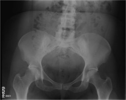
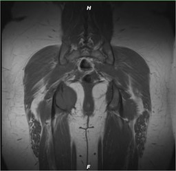
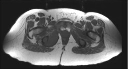
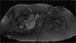
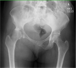
General Discussion:

The patient first presented to the ER with severe right hip pain radiating down her thigh. Plain films of the pelvis showed no abnormality. The patient subsequently underwent an MR which showed a 6.1 centimeter mass of the right ischium destroying the
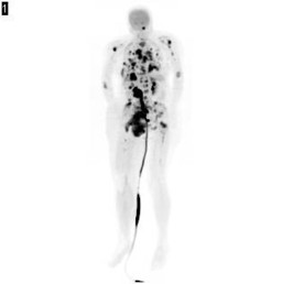
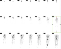
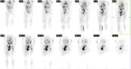
cortex. Subsequent imaging showed widespread metastatic disease. The patient was initiated on palliative radiation
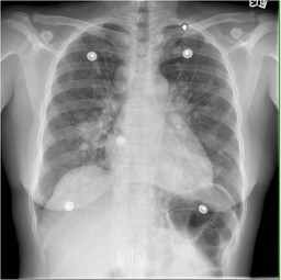
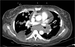
therapy. She was also treated for newly diagnosed large bilateral pulmonary emboli, as well as, a hospital-acquired pneumonia. Despite therapy, her cancer was highly aggressive.The patient expired five months after presenting to the ER with hip pain.

 Melanoma is a malignancy of pigment-producing cells (melanocytes) located predominantly in the skin, but also found in the eyes, ears, GI tract, leptomeninges, and oral and genital mucous membranes.  Malignant melanoma accounts for 4% of all skin cancers, but it causes 79% of skin cancer deaths. The incidence of melanoma has more than tripled in the white population during the last 20 years, and melanoma currently is the sixth most common cancer in the United States

Malignant melanoma is not a common cause of cancer metastasis to the skeleton. However, when melanoma does metastasize, one of the most common sites is the skeleton.

Melanoma is categorized in four stages by the American Joint Committee on Cancer.  Stage I has a Breslow depth of less than 2.01 mm and no ulceration; stage II has a Breslow depth of greater than 2.0 mm or lesions at 1.01 - 2.0 mm in depth with ulceration; stage III has regional lymph node metastasis; and stage IV has distant metastasis, which may include metastasis to bone. Autopsy studies indicate 23-49% osseus inlvolvement in patients who die from malignat melanoma. The mean survival time for patients with skeletal metastasis from malignant melanoma ranges from 3.6-4.7 months

Osseous metastases may remain invisible on plain films until cortical destruction has occurred, and scintigraphy may miss those lesions that are growing either too aggressively or too slowly to incite significant reactive bone formation.

References:

http://www.ajronline.org/cgi/reprint/155/1/109.pdf, Wd RH, Kaiser LR. Skeletal metastases of melanoma:


http://emedicine.medscape.com/article/1100753-overview#showall

http://www.bonetumor.org/metastatic-tumors/malignant-melanoma-metastasis-bone


radiographic, scintigraphic, and inical review.AJR 1981;137: 103-1 08


Comments:
No comments posted.
Additional Details:

Case Number: 323940Owner(s): Keith Fischer and Archana KantawalaLast Updated: 02-04-2013
Anatomy: Other   Pathology: Neoplasm
Modality: CT, Conventional Radiograph, MR, Nuc Med, PETAccess Level: Readable and writable by Nuclear Medicine only
Keywords: bsnm, psnm

Case has been viewed 9 times.
Certified by Keith Fischer on 02-04-2013

The reader is fully responsible for confirming the accuracy of this content.
Text and images may be copyrighted by the case author or institution.