Return to Case List with Diagnoses or Case List as Unknowns

HOT AND COLD LIVER METASTASIS ON SOMATOSTATIN RECEPTOR SCINTIGRAPHY
Authored By: kwinkl01 and Jonathan McConathy.
Patient: 63 year old female
History: 63 year old woman with rectal bleeding and abdominal pain.
Image Size:[small][as-submitted]

Multimedia: 284918_1_submitted.avi
SPECT Octreoscan

Fig. 2
SPECT Octreoscan

Fig. 3
SPECT Octreoscan

Fig. 4
SPECT Octreoscan

Fig. 5
SPECT Octreoscan

Fig. 6
Computed Tomography

Fig. 7
Computed Tomography

Fig. 8
Computed Tomography

Fig. 9
Computed Tomography
Image Size:[small][as-submitted]

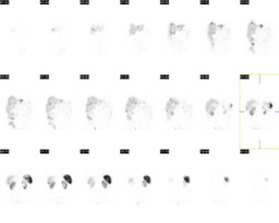
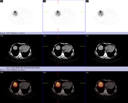
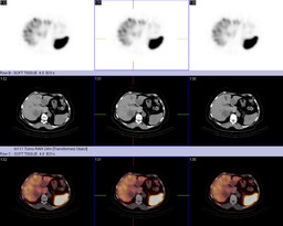
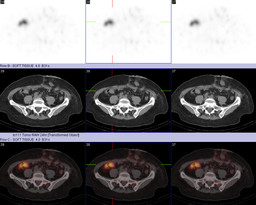
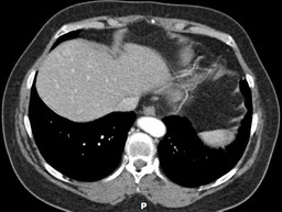
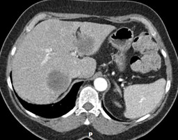
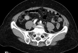
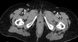
Findings:

SOMATOSTATIN-RECEPTOR SCINTIGRAPHY (WITH TOMOGRAPHIC IMAGING)

RADIOPHARMACEUTICAL:  6.0 mCi In-111 pentetreotide i.v.


There are two distinct populations of liver lesions. There are multiple low attenuation hepatic lesions with increased tracer accumulation. There are two large low-attenuation lesions which have less tracer accumulation than normal liver parenchyma.


There is an approximately 2.5 x 2.0 cm soft tissue density mass in the terminal ileum at the ileocecal valve with increased tracer uptake.


COMPUTED TOMOGRAPHY OF THE ABDOMEN AND PELVIS

There are multiple lesions seen throughout the liver.  Some of the hepatic lesions demonstrate hyperenhancement, while others demonstrate hypoenhancement.


There is a 2.4 cm hyperenhancing mass at the ileocecal valve.


There is eccentric wall thickening of the rectum, anteriorly concerning for rectal mass.  This mass is measures approximately 2 cm in axial dimension and 3 cm craniocaudally.  This mass is seen approximately 3 cm superior to the anal verge.

Diagnosis:

Well differentiated rectal adenocarcinoma with hepatic metastasis.


Well differentiated neuroendocrine carcinoma hepatic metastasis, with a probable terminal ileum primary.

General Discussion:

HISTORY

63 year old woman initially presented with rectal bleeding and abdominal pain.  Initial evaluation included abdominal sonography, which demonstrated several hepatic masses, as well as colonoscopy, which demonstrated a well differentiated rectal adenocarcinoma.  One of the hepatic masses was biopsied, which demonstrated a well differentiated neuroendocrine tumor.


Subsequent computed tomographic imaging and somatostatin receptor scintigraphy, demonstrated two distinct populations of liver lesions. There were multiple low attenuation hepatic lesions with increased tracer accumulation, consistent with neuroendocrine hepatic metastases, which correspond to hyperenhancing lesions on the contrast enhanced CT examination. There were two large low-attenuation lesions which had less tracer accumulation than normal liver parenchyma, which correspond to hypoenhancing lesions on the contrast enhanced CT examination.  These studies also demonstrated a soft tissue mass, within the terminal ileum with increased tracer accumulation, which correspond to a hyperenhancing mass on the contrast enhanced CT examination.  This likely represents the primary neuroendocrine tumor.  Subsequent liver biopsy demonstrated well differentiated adenocarcinoma metastasis.


DISCUSSION

Octreotide scintigraphy is based on neuropeptide receptor imaging.  Somatostatin is a neuropolypeptide and somatostatin receptors are present in an increased density within several neuroendocrine tumors.  Neuroendocrine tumors include carcinoid, pancreatic islet cell neoplasms, pheochromocytomas, neuroblastomas, paragangliomas, pituitary adenomas, medullary carcinoma of the thyroid, and small-cell lung cancers.  While many of these lesions are well evaluated with octreotide scintigraphy, exceptions include insulinomas and medullary thyroid carcinoma.  Planar imaging enables visualization of the entire body, which can be useful in locating the primary neoplasm, as well as metastatic disease.  Single photon emission computed tomography - computed tomography can be useful for better localization.  Octreotide scintigraphy has a higher sensitivity for well differentiated tumors, secondary to increased somatostatin receptor density.  Therefore, visualization may indicate a more favorable diagnosis, given well differentiation, as well as patients are more likely to be candidates for octreotide therapy.

References: Mettler, Fred and Milton Guiberteau. Essentials of Nuclear Medicine Imaging. 5th ed. Philadelphia, PA: Saunders Elsevier, 2006, pp 333-335.
Comments:
No comments posted.
Additional Details:

Case Number: 284918Owner(s): kwinkl01 and Jonathan McConathyLast Updated: 12-07-2011
Anatomy: Gastrointestinal (GI)   Pathology: Neoplasm
Modality: CT, Nuc MedAccess Level: Readable by all users, writable by NucMed Certifiers
Keywords: otnm

Case has been viewed 15 times.
Certified by Jonathan McConathy on 04-20-2011

The reader is fully responsible for confirming the accuracy of this content.
Text and images may be copyrighted by the case author or institution.