|
| Patient: 74 year old female |
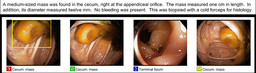
| History: 74 year-old woman with recurrent hypercalcemia and a presumed paraneoplastic syndrome.
Figure 1 Figure 2 Figure 3 Figure 4
Findings, differential, and Recommendations?
CT Images
Figure 5
Next step in management?
Figure 6 |
Image Size:
|
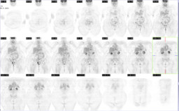
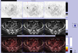
| Findings: PET-CT:
RADIOPHARMACEUTICAL: 14.8 mCi F-18 Fluorodeoxyglucose (FDG) i.v.
Focal moderate to markedly increased FDG uptake in the proximal cecum at the junction of the appendix and cecum. This is a nonspecific finding and can be seen in benign and malignant colonic lesions. Colonscopy is recommended.
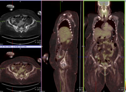
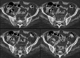
CT:
No definite abnormality noted in the proximal cecum at the junction of the appendix and the cecum.
Colonscopy:
Medium-sized mass in the cecum, right at the appendiceal orifice measuring 1 cm in length and 1.2 cm in diameter. See the colonoscopy pictures. |
| DDx: Single focus of increased FDG bowel uptake:
Malignant
Metastatsis
Primary colon cancer
Premalignant
adenomas
Benign
Hyperplastic Polyps
Infection/Inflammation |
| Diagnosis: Sessile serrated adenoma |
| References: Kei at al. "Incidental Finding of Focal FDG Uptake in the Bowel During PET/CT: CT Features and Correlation With Histopathologic Results" AJR 2010; 194: W401-W406. |
| Comments: No comments posted. |
| Additional Details:
Case Number: 245486 The reader is fully responsible for confirming the accuracy of this content. |