Return to Case List with Diagnoses or Case List as Unknowns

RIGHT UPPER LOBE FUNGAL INFECTION
Authored By: Farrokh Dehdashti and Collin Liu.
Patient: 65 year old female
History: 65 year old female: with metastatic leiomyosarcoma status post resection of the left retroperitoneal primary lesion, radiation therapy, and chemotherapy, currently on experimental chemotherapy.
Image Size:[small][as-submitted]

Fig. 1

Fig. 2
PET-CT

Fig. 3
PET-CT

Fig. 4
PET-CT

Fig. 5
PET-CT

Fig. 6
PET-CT

Fig. 7
Chest CT (2 months before the PET-CT)
Image Size:[small][as-submitted]

Findings:

PET-CT:
Radiopharmceutical: 9.9 mCi F-18 Fluorodeoxyglucose i.v.
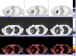
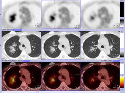
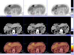
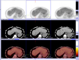
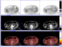
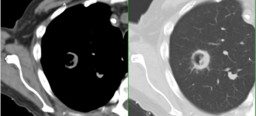
There has been significant interval increase in size of the right upper lobe cavitary lesion. This demonstrates a thick, intensely FDG-avid rim with surrounding groundglass attenuation and nodular consolidation (Figure 2). The maximal SUV is increased from 4.4 to 6.4 (prior PET-CT not shown). This is most consistent with a progressive infectious process such as a fungal infection. New nodular groundglass opacities are noted in the right perihilar lung (Figure 3). These demonstrate mild FDG uptake and likely represent spread of infection. Small nodules within the left upper lobe along the major fissure and in the right base have decreased in size when compared with the prior study. No associated FDG uptake is appreciated in these nodules; however, these are below the resolution of PET.

There are multiple low attenuation lesions in the peripheral aspect of segment 4B of the liver. The largest lesion has unchanged FDG uptake (Figure 4); however, it has decreased slightly in size compared with the prior study. The remaining lesions demonstrate FDG uptake comparable to the surrounding liver (not shown). There is unchanged, persistent intense FDG uptake in the region of the liver hilum (Figure 4). This may be within the liver, within a hilar lymph node, or within the duodenum.

A lobulated peritoneal mass in the left upper quadrant is unchanged in size and degree of mild FDG uptake (Figure 5 and Figure 6). Again seen is a low attenuation lesion in the spleen which shows mildly greater FDG uptake in the surrounding spleen, also unchanged (Figure 5).

Noncontrast CT images demonstrate postsurgical changes of left nephrectomy and partial colectomy with colocolonic anastomosis. Embolization coils are seen in the right hepatic and gastroduodenal arteries. There is associated atrophy of the right hepatic lobe. There is high attenuation material within low attenuation hepatic lesions in the liver dome and in segment 8, consistent with prior embolization.

Chest CT (2 months before PET-CT):
There has a new 2 cm cavitary lesion with asymmetric thick wall in the right upper lobe, with a small amount of surrounding ground glass opacity (Figure 7). There are multiple small pulmonary nodules consistent with the patient's known metastatic disease, which have all decreased in size compared to the prior study (not shown).


DDx: Cavitary metastasis, tuberculoma, primary bronchogenic tumor, bronchoalveolar carcinoma
Diagnosis:

Aspergiloma in the lung

General Discussion:

Full Patient History:
The patient is a 65-year-old female with left retroperitoneal leiomyosarcoma and metastasis to the liver, peritoneum, and spleen, status post resection of the left retroperitoneal primary lesion with associated left nephrectomy and left hemicolectomy, as well as chemoradiation therapy. She has also undergone radio frequency ventilation and chemoembolization of multiple liver lesions. At the time of the study, she was on experimental chemotherapy (Dacarbazine), admitted for malaise and elevated white blood cells, without fever.

General Discussion:
Aspergillosis can sometimes be seen in patients with impairment of immune system and/or preexisting lung disease (tuberculosis, bronchiectasis). It often mimicks malignancy on FDG-PET and CT, such as primary bronchogenic tumor or metastatic lesion. In this case, the rapid progression of the lesion over two months, and the associate infiltrative disease suggest an infectious etiology. The patient received right upper lobectomy after a bronchoscopic biopsy showed no malignancy. Aspergillus was identify in the mass.


References:

Invasive aspergillosis mimicking stage IIIA non-small-cell lung cancer on FDG positron emission tomography. Wilkinson MD, Fulham MJ, McCaughan BC, Constable CJ. Clin Nucl Med. 2003 Mar;28(3):234-5.

Radiology Review Manual, 6th Edition, Wolfgang Dahnert, MD, Page 465

Comments:
No comments posted.
Additional Details:

Case Number: 244138Owner(s): Farrokh Dehdashti and Collin LiuLast Updated: 12-08-2011
Anatomy: Cardiopulmonary   Pathology: Infection
Modality: CT, PETAccess Level: Readable and writable by Nuclear Medicine only
Keywords: fungal infection, pulmonary infection, ptnmACR: 60000.21700

Case has been viewed 15 times.
Certified by Farrokh Dehdashti on 12-08-2011

The reader is fully responsible for confirming the accuracy of this content.
Text and images may be copyrighted by the case author or institution.