Return to Case List with Diagnoses or Case List as Unknowns

SEPTIC ARTHRITIS OF THE RIGHT SACROILIAC JOINT ON PET IN A LYMPHOMA PATIENT
Authored By: Farrokh Dehdashti and Collin Liu.
Patient: 26 year old male
History:

26 year-old male with T-cell lymphoma, status post 3 cycles of chemotherapy, presents with lower back pain.


Image Size:[small][as-submitted]

Fig. 1
PET-CT #1

Fig. 2
PET-CT #1

Fig. 3
PET-CT #1

Fig. 4
PET-CT #2: 3 months later (after treatment)

Fig. 5
MRI TSE T2 FS (at the time of PET-CT #1)
Image Size:[small][as-submitted]

Findings:

PET-CT #1:
Radiopharmceutical: 14.8 mCi F-18 Fluorodeoxyglucose (FDG) i.v.
Linear increased FDG activity along the right sacroiliac joint without corresponding lytic or sclerotic bone lesions may reflect arthritis, an inflammatory or an infectious process (Figure 1 and Figure 2).

There is no abnormal FDG uptake to suggest recurrent or residual lymphoma. There is an anterior mediastinal mass with FDG uptake less than blood pool, representing treated lymphoma (Figure 3).


PET-CT #2 (post antibiotic treatment):
There is more intense linear FDG uptake along the course of the right sacroiliac joint compared to PET-CT #1, indicating active disease (Figure 4). There is resorption along the sacral aspect of the right sacroiliac joint.

MRI of the pelvis with contrast:
There is enhancing synovitis in the right sacroiliac joint with adjacent bone marrow edema in the sacrum and ilium. There is adjacent regional enhancement and edema within the right iliacus, obturator internus, iliopsoas, and gluteus musculature. This most likely represents septic arthritis (Figure 5).

DDx: Degenerative sacroiliitis, inflammatory spondyloarthropathies (including ankylosing spondylitis and arthritis associated with psoriasis), traumatic sacroiliitis
Diagnosis: Septic sacroiliitis
General Discussion:

Full Patient History:
The patient is a 26 year old male with T-cell lymphoma, status post 3 cycles of chemotherapy. He developed lower back pain, and sacroilitis was found on PET-CT #1. Subsequent biopsy and culture showed coagulase negative Staphylococcus septic arthritis. He was treated with intravenous antibiotics.

General Discussion:
Infection of the sacroiliac joint is extremely rare; fewer than 200 confirmed cases are reported in the English language literature. Its initial manifestations always mimic those of more common conditions, including low back pain, ruptured disk with sciatica, intra- or extra-pelvic abscess, psoas abscess, hip sepsis, abdominal infection, and pyelonephritis or kidney stones.

The etiology of this infection is generally accepted as the hematogenous spread of an organism to the highly vascular anterior sacroiliac region; in about half the cases, a remote, preexisting infection can be identified as the source of the responsible bacterium. Although this problem occurs among all age groups (with a possible 2:1 bias toward male patients), the majority of cases seem to occur in children and young adults. Staphylococcus aureus is the most frequently reported causative organism, but other infections have involved Streptococcus sp, Bacillus

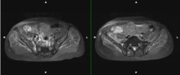
Early in the clinical course of this infection, pelvic radiographs almost always yield negative results; before the 1990s, the most reliable and recommended study for the early diagnosis of septic sacroiliitis was a bone scan. More recently, however, several reports have shown MRI of the pelvis to be far more sensitive and specific than nuclear medicine studies in rendering a correct diagnosis of sacroiliac infection

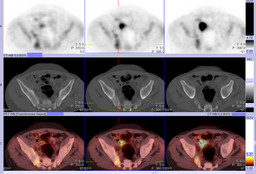
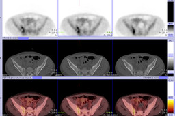
In this case, sacroiliitis was found incidentally on a PET-CT requested for monitoring treatment response of lymphoma. The linear pattern of FDG uptake in the right sacroiliac joint, without bone erosion, is suggestive of joint inflammation and/or infection. Subsequent pelvic MRI is supportive of this diagnosis. Joint aspiration and culture confirmed the diagnosis of septic sacroiliitis. Increased FDG uptake in the right sacroiliac joint seen on PET-CT #2 after weeks of intravenous antibiotic treatment likely indicates active disease.


References: 1. Hodgson BF: Pyogenic sacroiliac joint infection. Clin Orthop 246:146-149, 1989
2. Aprin H, Turen C: Pyogenic sacroiliitis in children. Clin Orthop 287:98-106, 1993
3. Abbott GT, Carty H: Pyogenic sacroiliitis, the missed diagnosis? Br J Radiol 66:120-122, 1993
Comments:
No comments posted.
Additional Details:

Case Number: 244112Owner(s): Farrokh Dehdashti and Collin LiuLast Updated: 12-08-2011
Anatomy: Skeletal System   Pathology: Infection
Modality: CT, MR, PETAccess Level: Readable and writable by Nuclear Medicine only
Keywords: septic arthritis, infection, inflammation, ptnmACR: 40000.26000

Case has been viewed 20 times.
Certified by Farrokh Dehdashti on 12-08-2011

The reader is fully responsible for confirming the accuracy of this content.
Text and images may be copyrighted by the case author or institution.