Return to Case List with Diagnoses or Case List as Unknowns

ANGIOIMMUNOBLASTIC T-CELL LYMPHOMA WITH C.DIFFICILE COLITIS ON FOLLOW UP
Authored By: Bennett Greenspan and Andrew Homb.
Patient: male
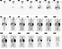
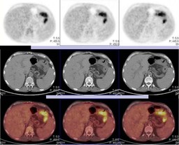
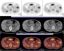
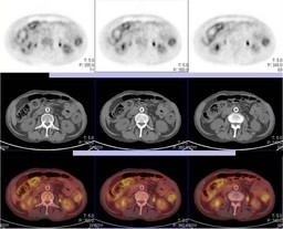
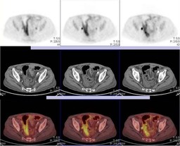
History:

65 year old man presents for restaging after completion of therapy


Figure 1

Figure 2

Figure 3

Figure 4

Figure 5

Figure 6

Figure 7


What is your interpretation of the study? 


The patient received an autologous stem cell transplant


One month after autologous stem cell transplantation


Figure 8

Figure 9

Figure 10

Figure 11

Figure 12

Figure 13

Figure 14


What is your interpretation of the follow up study?


Is there an important non-oncologic finding?

Image Size:[small][as-submitted]

Multimedia: 238067_1_submitted.avi
Rotating PET prior to stem cell transplant

Fig. 2
Coronal PET prior to stem cell transplant

Fig. 3
PET-CT prior to stem cell transplant

Fig. 4
PET-CT prior to stem cell transplant

Fig. 5
PET-CT prior to stem cell transplant

Fig. 6
PET-CT prior to stem cell transplant

Fig. 7
PET-CT prior to stem cell transplant

Multimedia: 238067_8_submitted.avi
Rotating PET 1 month after stem cell transplant

Fig. 9
Coronal PET 1 month after stem cell transplant

Fig. 10
PET-CT 1 month after stem cell transplant

Fig. 11
PET-CT 1 month after stem cell transplant

Fig. 12
PET-CT 1 month after stem cell transplant

Fig. 13
PET-CT 1 month after stem cell transplant

Fig. 14
PET-CT 1 month after stem cell transplant
Image Size:[small][as-submitted]

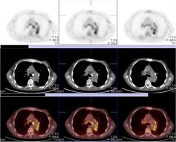
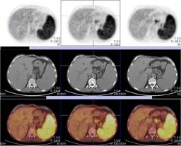
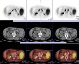
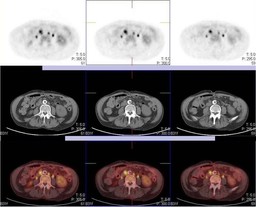
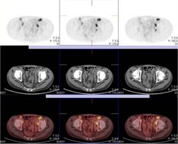
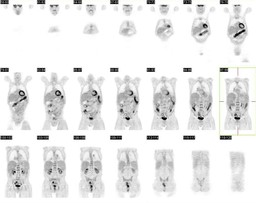
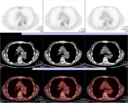
Findings:

Restaging after completion of therapy PET-CT Report


RADIOPHARMACEUTICAL: 12.4 mCi F-18 Fluorodeoxyglucose i.v.


1. Interval progression of disease with increased FDG uptake in cervical, mediastinal, hilar lymph, mesenteric, retroperitoneal and inguinal lymph nodes as well as abnormally increased FDG splenic uptake, increased compared to prior exam.


2. Stable left upper lobe spiculated nodule with blood pool level FDG uptake. Given the low uptake in the nodule relative to the patient's lymphadenopathy, this lesion is unlikely due to lymphoma. Continued imaging follow up is recommended as this nodule may represent a primary lung cancer such as bronchioloalveolar carcinoma.


One month after stem cell transplant PET-CT Report


RADIOPHARMACEUTICAL: 13.3 mCi F-18 Fluorodeoxyglucose (FDG) i.v.


1. Marked interval response to therapy with minimal to mild FDG uptake in hilar and subcarinal lymph nodes likely representing residual disease.


2. Interval development of diffusely increased colonic FDG uptake with wall thickening and inflammatory stranding.  


3. Interval development of multiple pulmonary nodules with minimal to mildly increased FDG uptake, likely inflammatory in etiology.


4. Spiculated left upper lobe pulmonary nodule with blood pool level FDG uptake, unchanged compared to prior exam. This nodule may represent a low grade primary lung cancer, and continued follow up is recommended.

DDx:

New diffusely increased colonic FDG uptake:


1. Infectious Colitis


2. Inflammatory Colitis


3. Atypical pattern of lymphomatous involvement of the colon

Diagnosis:

Diagnosis after restaging PET-CT:


Interval progression of angioblastic T-cell lymphoma (compared to a prior PET-CT, 3 months earlier)


Diagnosis one month after stem cell transplant:


Stool Specimen positive for Clostridium difficile toxin on 12/28/2009


General Discussion:

Full History:


65 year old man with angioimmunoblastic T-cell lymphoma presents for restaging after completion of therapy.  The second study was performed roughly a month after autologous stem cell transplant.  The patient reports persistent diarrhea since his stem cell transplant.


Discussion:


"Diarrhea is a major cause of morbidity and discomfort for patients undergoing high-dose chemotherapy with autologous peripheral blood stem cell transplantation (APBSCT) affecting 76-91% of transplant recipients."


Cause include:


Inflammation of the intestinal mucosa due to chemotherapy


Medications, including prophylactic antimicrobials


Infection (Most commonly C. difficile)


Arango et al. performed a retrospective study to determine the incidence of C. difficile-associated diarrhea (CDAD) 1 week prior to and 30 days after stem cell transplant and to identify any risk factors.


The study looked at 242 patients who underwent stem cell transplant for either lymphoma or myeloma.  Diarrhea was reported in 157 patients (64.9%).  The patients were all tested for C. difficle toxin.  The incidence of C. difficile in these 157 patients (1/3 lymphoma and 2/3 myeloma) was 15%.  The only significant indentifiable risk factors were the use of third generation cephalosporins and the use of intravenous vancomycin.  Three stool samples were need for diagnosis in most patients.  All patients responded to therapy with metronidazole or oral vancomycin.  

References: Arango et al. "Incidence of Clostridium difficile-associated diarrhea before and after autologous peripheral blood stem cell transplantation for lyphoma and multiple myeloma" Bone Marrow Transplantation. 37, 517-521. 2006
Comments:
No comments posted.
Additional Details:

Case Number: 238067Owner(s): Bennett Greenspan and Andrew HombLast Updated: 12-07-2011
Anatomy: Gastrointestinal (GI)   Pathology: Infection
Modality: Nuc Med, PETAccess Level: Readable by all users, writable by NucMed Certifiers
Keywords: ptnmACR: 90000.83000, 70000.26000

Case has been viewed 25 times.
Certified by Bennett Greenspan on 08-26-2011

The reader is fully responsible for confirming the accuracy of this content.
Text and images may be copyrighted by the case author or institution.