Return to Case List with Diagnoses or Case List as Unknowns

METASTATIC CERVICAL CANCER
Authored By: Peter Phan and Henry Royal.
Patient: 38 year old female
History: 38-year-old female with cervical cancer, diagnosed and treated two years ago.  She now presents with productive cough and fevers.
Image Size:[small][as-submitted]

Multimedia: 206896_1_submitted.avi
PET-CT

Fig. 2
PET-CT

Fig. 3
PET-CT

Fig. 4
Subsequent Contrast CT of the Chest
Image Size:[small][as-submitted]

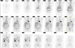
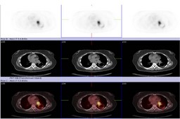
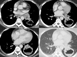
Findings: RADIOPHARMACEUTICAL: 14.6 mCi F-18 Fluorodeoxyglucose i.v.

With comparison to a prior PET-CT (interpreted as negative for malignancy) there has been interval development of bulky left hilar and AP window lymphadenopathy that are highly FDG avid. There is occlusion of the left main stem bronchus. Distal to this, in the left lower lobe, there is a 6 cm thick walled cavitary mass with adjacent ground glass opacities; the wall of this cavity is FDG avid. No abnormal FDG uptake within the abdomen or pelvis to suggest local recurrent malignant disease.
 
Diagnosis: Pulmonary metastasis from treated cervical squamous cell carcinoma
General Discussion: The patient was diagnosed with cervical cancer and treated two years ago.  The interval present of a cavitary mass and mediastinal adenopathy may represent primary lung cancer.  However, in this case, it is equally probable that this finding represents metastasis from cervical cancer, as it turned out to be.

The spread of cervical cancer is usually predictable, involving local lymphatic structures in a systematic  pattern, generally involving deep pelvic, para-aortic, and supraclavicular stations in that order.  Distant solid organ and pulmonary metastasis generally follows only after extensive nodal involvement.  However, this predictable pattern only applies to patients who have not had any treatment.  After the treatment of cervical cancer, the pattern of metastasis is not so predictable and subsequently, one can find distant pulmonary metastasis without pelvic nodal disease on restaging PET-CT exams.


Comments:
No comments posted.
Additional Details:

Case Number: 206896Owner(s): Peter Phan and Henry RoyalLast Updated: 12-07-2011
Anatomy: Genitourinary (GU)   Pathology: Neoplasm
Modality: PETAccess Level: Readable by all users, writable by NucMed Certifiers
Keywords: ptnm

Case has been viewed 44 times.
Certified by Henry Royal on 09-17-2009

The reader is fully responsible for confirming the accuracy of this content.
Text and images may be copyrighted by the case author or institution.