Return to Case List with Diagnoses or Case List as Unknowns

LYMPHOMA OF THE PERITONEUM
Authored By: Farrokh Dehdashti and Peter Phan.
Patient: 36 year old male
History: The patient is a 36 year old male with progressive abdominal distention.
Image Size:[small][as-submitted]

Multimedia: 206893_1_submitted.avi
PET/CT

Fig. 2
Coronal PET images

Fig. 3
Axial PET/CT images

Fig. 4
Axial PET/CT images

Fig. 5
Axial PET/CT images
Image Size:[small][as-submitted]

Findings: There is diffuse omental nodularity and mesenteric adenopathy that is highly FDG avid. Hypermetabolic soft tissue is present within the right anterior mediastinum extending along the right pericardium, to the right cardiophrenic space and diaphragmatic surface.
Diagnosis: Non-Hodgkin Burkitt's lymphoma with peritoneal lymphomatosis and mediastinal involvement.
General Discussion: RADIOPHARMACEUTICAL: 15.0 mCi F-18 Fluorodeoxyglucose i.v.

Metastatic ovarian cancer is generally the usual suspect when isolated peritoneal carcinomatosis is found on CT.  However, in this male patient, other entities such as metastasis from a gastrointestinal malignancy, or a primary tumor of the peritoneum were considered.   

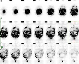
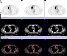
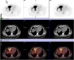
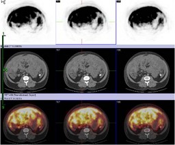
Peritoneal lymphomatosis is the most likely diagnosis in this case because of the intense degree of FDG uptake and the presence of mediastinal adenopathy.  Isolated peritoneal lymphomatosis is a rare manifestation of non-Hodgkin lymphoma, but is an uncommon finding when other sites of disease are present. 

The differential diagnosis of peritoneal carcinomatosis can be divided into two broad categories: metastatic disease and primary tumor of the peritoneum.  While metastatic disease is by far more common (generally from the gastrointestinal tract and ovaries), primary peritoneal neoplasm should be considered when a known primary is not clearly evident.


Primary peritoneal neoplasm includes malignant tumors such as malignant mesothelioma, papillary serous carcinoma, desmoplastic small round cell tumor, lymphoproliferative disorders (peritoneal lymphomatosis and leukemic infiltration), as well as mesenchymal tumors (liposarcoma, angiosarcoma, malignant fibrous histiocytoma).


Considering the hypermetabolic nature of the findings, a primary benign peritoneal tumor is unlikely in this case.  This includes cystic mesothelioma and benign mesenchymal tumors (lymphangioma, hemangioma, leiomyomatosis peritonealis).

References: 1.  Perry J. Pickhardt and Sanjeev Bhalla. "Primary Neoplasms of Peritoneal and Sub-peritoneal Origin: CT Findings" RadioGraphics 2005; 25: 983-995.

2. Even-Sapir E, Lievshitz G, Perry C, Herishanu Y, Lerman H, Metser U. Fluorine-18 fluorodeoxyglucose PET/CT patterns of extranodal involvement in patients with Non-Hodgkin lymphoma and Hodgkin's disease. Radiol Clin North Am. 2007; 45:697-709.
Comments:
No comments posted.
Additional Details:

Case Number: 206893Owner(s): Farrokh Dehdashti and Peter PhanLast Updated: 12-07-2011
Anatomy: Gastrointestinal (GI)   Pathology: Neoplasm
Modality: Nuc Med, PETAccess Level: Readable by all users, writable by NucMed Certifiers
Keywords: ptnmACR: 70000.34000

Case has been viewed 39 times.
Certified by Farrokh Dehdashti on 07-07-2008

The reader is fully responsible for confirming the accuracy of this content.
Text and images may be copyrighted by the case author or institution.