
 |
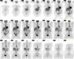
| Fig. 1 |
| Coronal whole body PET images demonstrate no gross abnormalities |
|
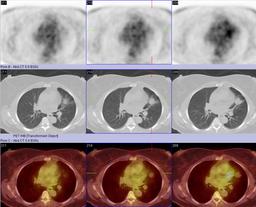 |
| Fig. 2 |
| PET, CT, and fused images demonstrating a lingular mass with mild tracer uptake. |
|
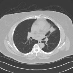 |
| Fig. 3 |
| Non-contrast CT image in lung windows demonstrates spiculated lingular mass |
|
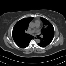 |
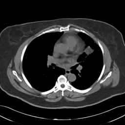
| Fig. 4 |
| Non-contrast axial CT image demonstrating lingular mass containing fat. |
|
 |
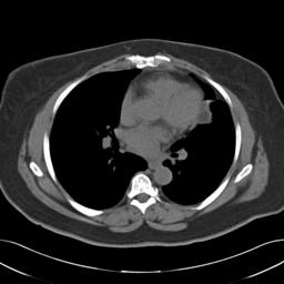
| Fig. 5 |
| Non-contrast axial CT image demonstrating lingular mass containing fat. |
|
 |
| Fig. 6 |
| Non-contrast axial CT image demonstrating lingular mass containing fat. |
|
 |
| Fig. 7 |
| Non-contrast axial CT image demonstrating lingular mass containing fat. |
|