Return to Case List with Diagnoses or Case List as Unknowns

LARGE B-CELL LYMPHOMA
Authored By: Asif Moinuddin and Henry Royal.
Patient: 47 year old female
History: 47-year-old woman with large B-cell lymphoma.
Image Size:[small][as-submitted]

Fig. 1
Initial images

Fig. 2
Initial images

Fig. 3
Initial images

Fig. 4
Five months later

Fig. 5
Five months later
Image Size:[small][as-submitted]

Findings: RADIOPHARMACEUTICAL: 14 mCi F-18 Fluorodeoxyglucose i.v.
FINDINGS:
Mild FDG-avid soft tissue mass in the region of the right nasal fold, anterior to the right maxillary sinus. Given the biopsy result of a recently excised adjacent right cheek mass, this most likely represents lymphoma. Conglomeration of mesenteric lymph nodes measuring 4.1 x 3.7 cm in greatest dimension with no FDG uptake and multiple enlarged submandibular, single right supraclavicular, and several mesenteric lymph nodes with no FDG uptake. However with recently diagnosed lymphoma in the right cheek mass, this pattern of lymph node involvement raises the suspiscion of lymphoma at these sites.
General Discussion: FOLLOW-UP:
Follow-up scan after 6 cycles of R-CHOP demonstrated near complete resolution of FDG uptake. 3 months later, a restaging PET scan was negative for tumor involvement (images not shown)
References:

Weihrauch et al; Ann Hem 2002 81: 20-25
Jerusalem et al: hematologica 2000  85  613-618
Mikosch P et al; Acta Medica Austriaca: 2003;Heft 2, 41-47 

 20-25

Comments:
No comments posted.
Additional Details:

Case Number: 206890Owner(s): Asif Moinuddin and Henry RoyalLast Updated: 12-07-2011
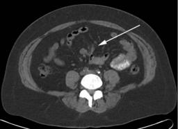
Anatomy: Vascular/Lymphatic   Pathology: Neoplasm
Modality: Nuc Med, PETAccess Level: Readable by all users, writable by NucMed Certifiers
Keywords: ptnm, lymphoma, hidgkin disease, non-hodgkin lymphoma, sarcoma

Case has been viewed 27 times.
Certified by Henry Royal on 10-15-2010

The reader is fully responsible for confirming the accuracy of this content.
Text and images may be copyrighted by the case author or institution.