|
| Patient: 51 year old male |
| History: 51-year-old man with left shoulder melanoma. The patient initially presented approximately 5-6 years ago with a cutaneous lesion on the lateral aspect of his left shoulder, overlying the deltoid. The lesion was excised and per the patient, was found to be benign on pathologic examination. In December, 2006 the patient found a new reddish-colored lesion in that same area. This lesion was excised on February 23, 2007 and found to be malignant melanoma. Perform PET/CT for initial staging of disease. |
Image Size:
|
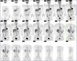
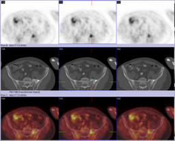
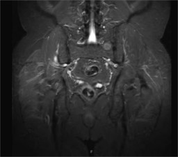
| Findings: The FDG-PET images demonstrate a single focus of abnormal FDG uptake located within the left sacral ala. There is no underlying lesion demonstrated on the correlative CT bone windows. Given the focal uptake, this is concerning for osseous metastatic disease. |
| Diagnosis: Fibrous dysplasia confirmed by biopsy. |
| General Discussion: The very focal nature of the intense uptake without an underlying radiographic abnormality made it concerning for a metastasis. |
| References: Radiology. 2001;219:774-777 Skeletal Radiol. 2007 Jun;36 Suppl 1:S24-8. Epub 2006 May 20. |
| Comments: No comments posted. |
| Additional Details:
Case Number: 206887 The reader is fully responsible for confirming the accuracy of this content. |