Return to Case List with Diagnoses or Case List as Unknowns

ELASTOFIBROMA
Authored By: Farrokh Dehdashti and O'Neil Lee.
Patient: 66 year old female
History: 63 year old female with history of endometrical carcinoma and renal cell carcinoma status post surgery and chemotherapy. Now presents with elevated CA-125 and negative CT scan.
Image Size:[small][as-submitted]

Fig. 1

Fig. 2

Fig. 3
Image Size:[small][as-submitted]

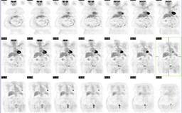
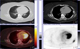
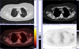
Findings: 1. Increased FDG uptake within a soft tissue density between the left scapula and chest wall just inferior to the scapula (Figure 2).

2. Increased FDG uptake within the left axillary lymph node (Figure 3).
Diagnosis: Elastofibroma.
General Discussion: Elastofibroma is a benign connective tussue tumor mostly seen in middle-aged women.  Their characteristic location (usually located between the scapula and chest wall along the inferior aspect of the scapula) is helpful in diagnosis.  Classic CT/MRI appearance is a soft tissue mass with interspersed strands of fat (as seen in this case).  Increased FDG uptake has been reported with elastofibroma (Reference 1). 

Increased uptake in the left axillary lymph node was felt to be secondary to infiltration of the tracer in the antecubital fossa.  The lymph node had been stable in size over the past 1-2 years.
References: Pierce, JC. AJR 2004. 183:35-37.
Comments:
No comments posted.
Additional Details:

Case Number: 206884Owner(s): Farrokh Dehdashti and O'Neil LeeLast Updated: 12-07-2011
Anatomy: Skeletal System   Pathology: Benign Mass, Cyst
Modality: PETAccess Level: Readable by all users, writable by NucMed Certifiers
Keywords: elastofibromaACR: 40000.36000

Case has been viewed 30 times.
Certified by Farrokh Dehdashti on 07-07-2008

The reader is fully responsible for confirming the accuracy of this content.
Text and images may be copyrighted by the case author or institution.