Return to Case List with Diagnoses or Case List as Unknowns

DEMENTIA
Authored By: Xiaoni Hong and Tom Miller.
Patient: 74 year old male
History: 74-year-old man who was recently found to have a memory disorder.
Image Size:[small][as-submitted]

Fig. 1
Transaxial brain PET

Fig. 2
Sagittal brain PET

Fig. 3
Transaxial fusion images of brain PET/Ct

Fig. 4
Sagittal fusion images of brain PET/CT

Fig. 5
Coronal fusion imagings of brain PET/CT
Image Size:[small][as-submitted]

Findings:

FDG PET: There is decreased metabolism in both frontal lobes. There is also possible decreased metabolism in the anterior portion of both temporal lobes. There is normal metabolism in the parietal lobes, posterior portions of the temporal lobes and the occipital lobes.

On the non-contrast CT examination, there is atrophy of the frontal lobes and the anterior portion of the temporal lobes.

Diagnosis: Frontotemporal  dementia (Pick's disease).
General Discussion:

This 74-year-old man was recently found to have a memory disorder.

Discussion: Pick's disease is now referred to as frontotemporal dementia. The onset of the disease is typically in the mid-to-late 50's, and the disease averages about 10 years from onset to death. The cause remains unknown.  Before PET, the only way to diagnose frontotemporal dementia was at autopsy. Clinical symptoms include disturbances of personality, behavior and language which may be noticed first and even be more severe than memory defects. Depending on the lobe affected, symptoms may include loss of social and sexual inhibition, impaired judgment, hoarding items, roaming, difficulties with attention and motivation, aphasia, repetitive speech patterns or the tendency to repeat anything heard.
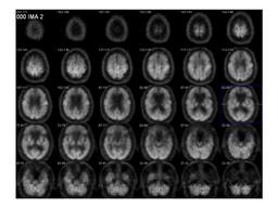
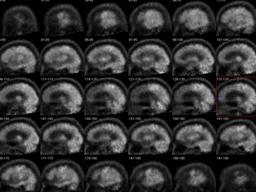
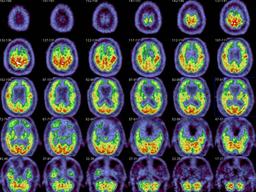
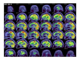
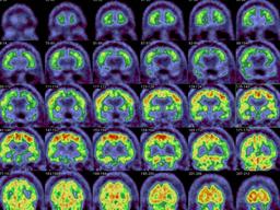

There is atrophy of the frontotemporal regions on CT or MRI. Decreased FDG uptake in the frontal lobes and anterior temporal lobes is a feature of frontotemporal dementia. This case demonstrates te classic pattern of hypometabolism in the frontotemporal lobes on PET/CT.

References: 1. Kazunari Ishii, Setsu Sakamoto, Masahiro Sasaki, et al. Cerebral Glucose Metabolism in Patients with Frontotemporal Dementia. J. Nucl. Med. 1998 39: 1875-1878.

2.Yong Jeong, Sang Soo Cho, Jung Mi Park, et al.18F-FDG PET Findings in Frontotemporal Dementia: An SPM Analysis of 29 Patients.J. Nucl. Med. 2005 46: 233-239. 

3.Kazunari Ishii, Setsu Sakamoto, Masahiro Sasaki, et al. Cerebral Glucose Metabolism in Patients with frontotemporal Dementia. J. Nucl. Med. 1998 39: 1875-1878. 
Comments:
No comments posted.
Additional Details:

Case Number: 206882Owner(s): Xiaoni Hong and Tom MillerLast Updated: 12-07-2011
Anatomy: Cranium and Contents   Pathology: Other
Modality: PETAccess Level: Readable by all users, writable by NucMed Certifiers
Keywords: pbnm, dementiaACR: 10000.83000

Case has been viewed 54 times.
Certified by Tom Miller on 04-18-2007

The reader is fully responsible for confirming the accuracy of this content.
Text and images may be copyrighted by the case author or institution.