|
| ||||||
|
|
|
| Patient: 54 year old |
| History: Presenting for evaluation of pulmonary nodules. |
Image Size:
|
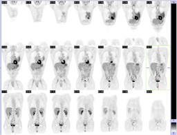
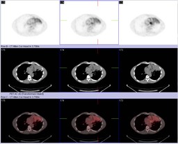
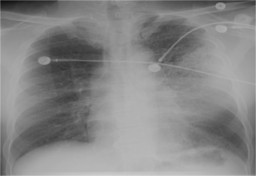
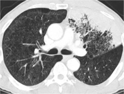
| Findings: The PET-CT images demonstrate focal, moderate FDG-avid consolidation in the left upper lobe (Fig. 1, Fig. 2).Ā There were additionally small pulmonary nodules, predominantly in the superior segment of the left lower lobe, too small to characterize (not shown). [pagebreak] The chest radiograph and CT obtained approximately 6 weeks later showed persistent left upper lobe consolidation (Fig. 3, Fig. 4).Ā The CT also re-demonstrated small left lower lobe (superior segment)Ānodules. |
| Diagnosis: Bronchoalveolar cell carcinoma |
| References: 1.Ā Yap CS et al, "FDG-PET imaging in lung cancer: how sensitive is it for bronchioloalveolar carcinoma?" Eur J Nucl Med 2002 Nov; 29(9): 1166-1173. 2.Ā Gandara DR, "Radiographic imaging of bronchioloalveolar carcinoma: screening, patterns of presentation and response assessment,"Ā J Thorac Oncol 2006 Nov;1(9 Suppl):S20-6. |
| Comments: No comments posted. |
| Additional Details:
Case Number: 206878 The reader is fully responsible for confirming the accuracy of this content. |