Return to Case List with Diagnoses or Case List as Unknowns

ADRENAL CARCINOMA ON PET
Authored By: O'Neil Lee and Henry Royal.
Patient: 57 year old female
History: 57 year old female with lower extremity edema.
Image Size:[small][as-submitted]

Fig. 1

Fig. 2

Fig. 3
Image Size:[small][as-submitted]

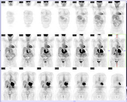
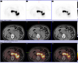
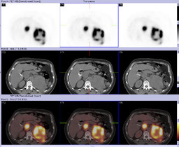
Findings: FDG-PET CT demonstrates a large left suprarenal mass with increased FDG uptake (and central photopenia).

Additionally, increased uptake is seen within the left renal vein and IVC.
Diagnosis: Adrenal carcinoma (biopsy proven).
General Discussion: Similar to renal cell carcinoma, adrenal carcinoma can invade into the renal vein and IVC.  This case nicely demonstrates venous invasion of the carcinoma with markedly increased FDG uptake within the adrenal mass (with central necrosis) and intravascular tumor.
Comments:
No comments posted.
Additional Details:

Case Number: 206876Owner(s): O'Neil Lee and Henry RoyalLast Updated: 12-07-2011
Anatomy: Genitourinary (GU)   Pathology: Neoplasm
Modality: PETAccess Level: Readable by all users, writable by NucMed Certifiers
Keywords: adrenal carcinoma

Case has been viewed 21 times.
Certified by Henry Royal on 09-17-2009

The reader is fully responsible for confirming the accuracy of this content.
Text and images may be copyrighted by the case author or institution.