|
| ||||||
|
| ||||||
|
|
|
| Patient: 74 year old |
| History: 74 year old patient: Recurrent squamous cell carcinoma of the right pharynx, on chemotherapy.Ā Monitoring treatment response. |
Image Size:
|
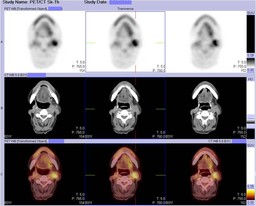
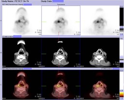
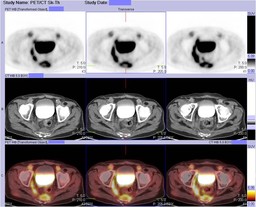
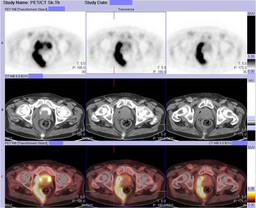
| Findings: Radiopharmaceutical:Ā 14 mCi F-18 Fluorodeoxyglucose i.v. The current rotating MIP images (Fig. 1) demonstrate two areas of focal uptake in the neck, improved since the prior examination (Fig. 2).Ā However, there is a new area of abnormal FDG activity in the right pelvis (Fig. 1). Axial PET-CT images confirm a metastatic lymph node in the left neck (Fig. 3), as well as uptake in the primary tumor in the rightĀsupraglottic larynx (Fig. 4).Ā Additionally, the focal uptake in the pelvis corresponds toĀgas-containing inflammatory soft tissueĀandĀfluidĀin the right pelvis and presacral space (Fig. 5), abutting a thickened rectal wall (Fig. 6). |
| Diagnosis: Right pelvic and presacralĀabscess, presumably due to a perforated anus or rectum. |
| Comments: No comments posted. |
| Additional Details:
Case Number: 174057 The reader is fully responsible for confirming the accuracy of this content. |