Return to Case List with Diagnoses or Case List as Unknowns

INCIDENTALLY IDENTIFIED COLON CANCER DURING PET-CT FOLLOW-UP OF LYMPHOMA TREATMENT
Authored By: Akash Sharma and Jonathan Weiss.
Patient: 66 year old female
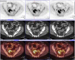
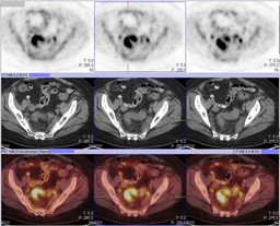
History:

66 year old female:

Patient has Non-Hodgkin's lymphoma involving the cervix.   She had an initial staging PET-CT (Figure 1) and a treatment monitoring PET-CT (Figure 2) two months later while she was receiving chemotherapy.

Image Size:[small][as-submitted]

Multimedia: 165135_1_submitted.avi
Initial staging PET-CT

Multimedia: 165135_2_submitted.avi
Treatment monitoring PET-CT

Fig. 3
PET-CT

Multimedia: 165135_4_submitted.avi
PET-CT

Fig. 5
PET-CT

Fig. 6
PET-CT
Image Size:[small][as-submitted]

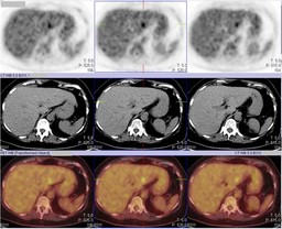
Findings:

RADIOPHARMACEUTICAL: 13.6 mCi F-18 Fluorodeoxyglucose i.v.

Patients initial staging PET-CT (Figure 1) shows multiple foci of increased FDG uptake involving the cervix, left uterus, internal iliac lymph nodes, presacral region, sacrum, T2 vertebral body and left iliac bone.  There is also a short segment of increased FDG uptake in the sigmoid colon.

Patients treatment monitoring PET-CT (Figure 2) shows a marked response to therapy as evidenced by the resolution of uptake in the uterus, cervix, presacral soft tissue, and internal iliac lymph nodes as well as in the left iliac bone and acetabulum with minimal residual uptake in the sacrum and T2 vertebral body.  However, there is persistent FDG activity within rectosigmoid colon (Figure 3).

DDx:

Physiologic bowel uptake

Colon cancer

Inflammatory bowel disease

Infectious colitis

Diagnosis:

Adenocarcinoma of the rectosigmoid colon

General Discussion:

Patient subsequently had a colonoscopy and biopsy of the rectosigmoid colon.  Patient then had an initial staging PET-CT for colon cancer that showed an intense focal area of uptake in the liver that was most consistent with metastatic disease (Figure 4 and Figure 5).

The colon can have variable FDG uptake.  Typically the cecum and right colon have relatively higher FDG uptake possibly due to higher concentrations of lymphocytes.  Focal or segmental FDG uptake in the colon must be carefully evaluated.  Differential diagnosis includes a benign or malignant tumor, inflammatory bowel disease, infectous colitis and physiologic bowel uptake.  Correlation with CT images and the pattern of FDG uptake is often helpful.  In this case, the segmental uptake that was persistent after treatment, especially with the improvement of disease elsewhere, was very suspicious for a second primary malignancy.  Focal increased FDG uptake in the colon on a PET-CT should be further evaluated with colonoscopy to exclude an underlying lesion.

References:

Hima B. Prabhakar, Dushyant V. Sahani, Alan J. Fischman, Peter R. Mueller, and Michael A. Blake.  Bowel Hot Spots at PET-CT.  RadioGraphics 2007 27: 145-159.

Comments:
No comments posted.
Additional Details:

Case Number: 165135Owner(s): Akash Sharma and Jonathan WeissLast Updated: 12-07-2011
Anatomy: Gastrointestinal (GI)   Pathology: Neoplasm
Modality: PETAccess Level: Readable by all users, writable by NucMed Certifiers
Keywords: ptnmACR: 00007.32000

Case has been viewed 47 times.

The reader is fully responsible for confirming the accuracy of this content.
Text and images may be copyrighted by the case author or institution.